4H51 image
Deposition Date 2012-09-18
Release Date 2012-10-10
Last Version Date 2025-03-26
Entry Detail
PDB ID:
4H51
Keywords:
Title:
Crystal structure of a putative Aspartate Aminotransferase from Leishmania major Friedlin
Biological Source:
Source Organism:
Host Organism:
Method Details:
Experimental Method:
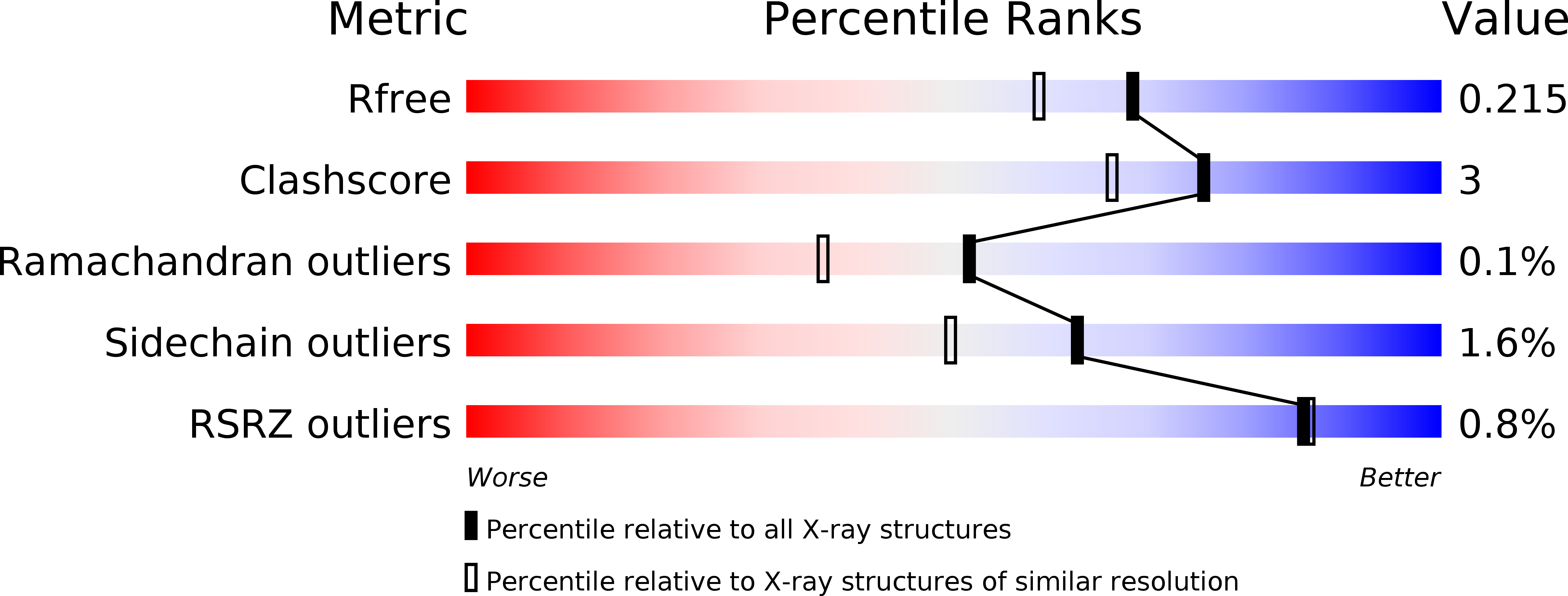
Resolution:
1.85 Å
R-Value Free:
0.21
R-Value Work:
0.16
R-Value Observed:
0.16
Space Group:
P 1 21 1
Macromolecular Entities
Structures with similar UniProt ID
Protein Blast
Polymer Type:polypeptide(L)
Molecule:Aspartate aminotransferase
Gene (Uniprot):asat
Chain IDs:A, B
Chain Length:420
Number of Molecules:2
Biological Source:Leishmania major
Modified Residue
Compound ID Chain ID Parent Comp ID Details 2D Image
LLP A LYS ?
Ligand Molecules
Primary Citation
Structures of aspartate aminotransferases from Trypanosoma brucei, Leishmania major and Giardia lamblia.
Acta Crystallogr F Struct Biol Commun 71 566 571 (2015)
PMID: 25945710 DOI: 10.1107/S2053230X15001831

Abstact

The structures of three aspartate aminotransferases (AATs) from eukaryotic pathogens were solved within the Seattle Structural Genomics Center for Infectious Disease (SSGCID). Both the open and closed conformations of AAT were observed. Pyridoxal phosphate was bound to the active site via a Schiff base to a conserved lysine. An active-site mutant showed that Trypanosoma brucei AAT still binds pyridoxal phosphate even in the absence of the tethering lysine. The structures highlight the challenges for the structure-based design of inhibitors targeting the active site, while showing options for inhibitor design targeting the N-terminal arm.

Legend

Protein

Chemical

Disease

Primary Citation of related structures
Feedback Form
Name
Email
Institute
Feedback