4H4F image
Deposition Date 2012-09-17
Release Date 2013-02-27
Last Version Date 2024-10-30
Entry Detail
PDB ID:
4H4F
Title:
Crystal structure of human chymotrypsin C (CTRC) bound to inhibitor eglin c from Hirudo medicinalis
Biological Source:
Source Organism:
Homo sapiens (Taxon ID: 9606)
Hirudo medicinalis (Taxon ID: 6421)
Method Details:
Experimental Method:
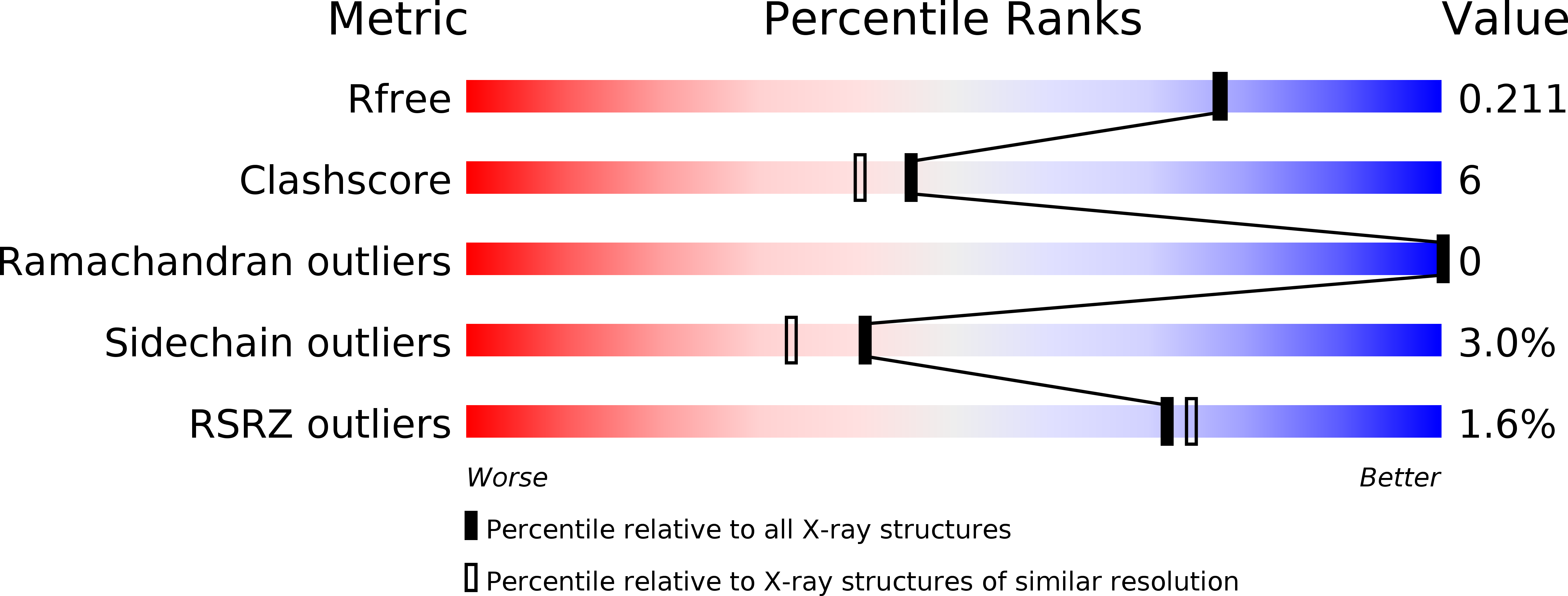
Resolution:
1.90 Å
R-Value Free:
0.20
R-Value Work:
0.15
R-Value Observed:
0.15
Space Group:
P 21 21 21
Macromolecular Entities
Polymer Type:polypeptide(L)
Molecule:Chymotrypsin-C
Gene (Uniprot):CTRC
Chain IDs:A
Chain Length:249
Number of Molecules:1
Biological Source:Homo sapiens
Polymer Type:polypeptide(L)
Molecule:Eglin C
Chain IDs:B
Chain Length:70
Number of Molecules:1
Biological Source:Hirudo medicinalis
Polymer Type:polypeptide(L)
Molecule:Chymotrypsin-C
Gene (Uniprot):CTRC
Chain IDs:C (auth: Q)
Chain Length:10
Number of Molecules:1
Biological Source:Homo sapiens
Ligand Molecules
Primary Citation
Long-range Electrostatic Complementarity Governs Substrate Recognition by Human Chymotrypsin C, a Key Regulator of Digestive Enzyme Activation.
J.Biol.Chem. 288 9848 9859 (2013)
PMID: 23430245 DOI: 10.1074/jbc.M113.457382

Abstact

Human chymotrypsin C (CTRC) is a pancreatic serine protease that regulates activation and degradation of trypsinogens and procarboxypeptidases by targeting specific cleavage sites within their zymogen precursors. In cleaving these regulatory sites, which are characterized by multiple flanking acidic residues, CTRC shows substrate specificity that is distinct from that of other isoforms of chymotrypsin and elastase. Here, we report the first crystal structure of active CTRC, determined at 1.9-Å resolution, revealing the structural basis for binding specificity. The structure shows human CTRC bound to the small protein protease inhibitor eglin c, which binds in a substrate-like manner filling the S6-S5' subsites of the substrate binding cleft. Significant binding affinity derives from burial of preferred hydrophobic residues at the P1, P4, and P2' positions of CTRC, although acidic P2' residues can also be accommodated by formation of an interfacial salt bridge. Acidic residues may also be specifically accommodated in the P6 position. The most unique structural feature of CTRC is a ring of intense positive electrostatic surface potential surrounding the primarily hydrophobic substrate binding site. Our results indicate that long-range electrostatic attraction toward substrates of concentrated negative charge governs substrate discrimination, which explains CTRC selectivity in regulating active digestive enzyme levels.

Legend

Protein

Chemical

Disease

Primary Citation of related structures