4H3K image
Deposition Date 2012-09-13
Release Date 2012-10-31
Last Version Date 2024-10-30
Entry Detail
PDB ID:
4H3K
Keywords:
Title:
Crystal structure of a ternary complex of human symplekin NTD, human Ssu72 and a RNA polymerase II CTD peptide phosphorylated at Ser-2, Ser-5 and Ser-7
Biological Source:
Source Organism:
Homo sapiens (Taxon ID: 9606)
Host Organism:
Method Details:
Experimental Method:
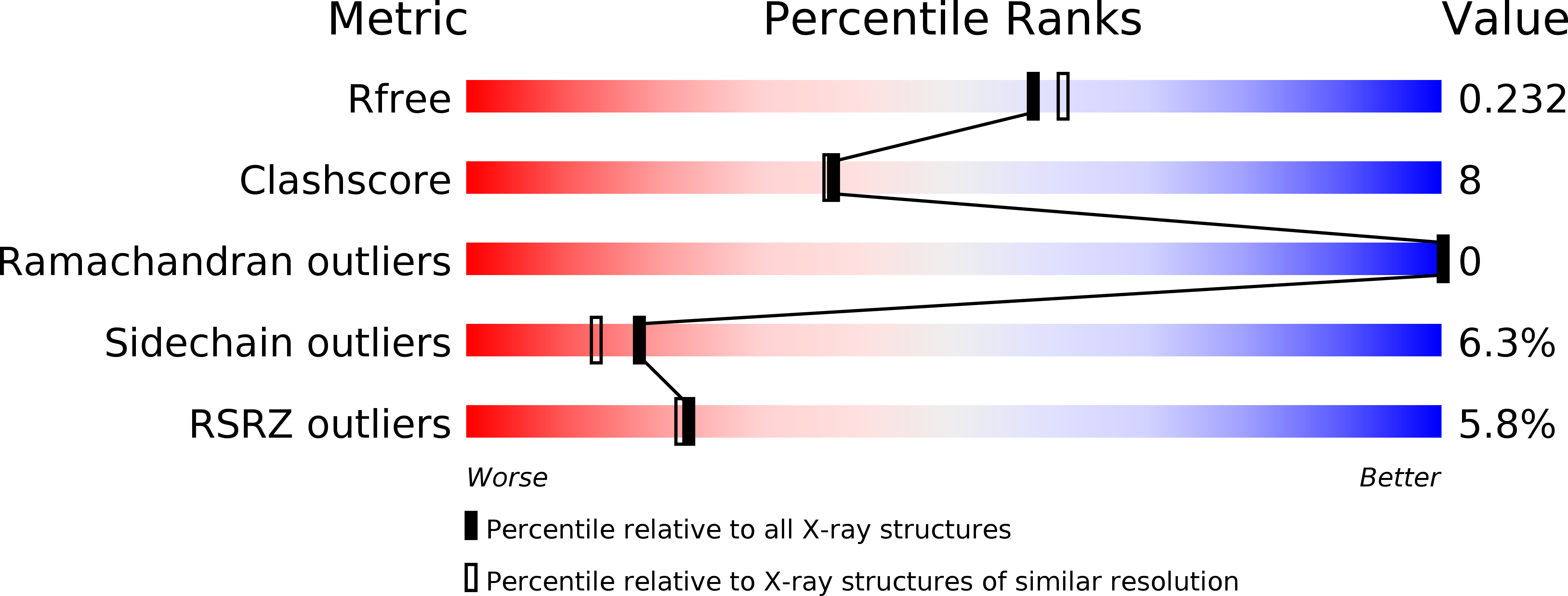
Resolution:
2.00 Å
R-Value Free:
0.23
R-Value Work:
0.19
R-Value Observed:
0.19
Space Group:
P 1 21 1
Macromolecular Entities
Structures with similar UniProt ID
Protein Blast
Polymer Type:polypeptide(L)
Molecule:Symplekin
Gene (Uniprot):SYMPK
Chain IDs:A, C (auth: D)
Chain Length:351
Number of Molecules:2
Biological Source:Homo sapiens
Structures with similar UniProt ID
Protein Blast
Polymer Type:polypeptide(L)
Molecule:RNA polymerase II subunit A C-terminal domain phosphatase SSU72
Gene (Uniprot):SSU72
Mutagens:C12S
Chain IDs:B, D (auth: E)
Chain Length:214
Number of Molecules:2
Biological Source:Homo sapiens
Modified Residue
Compound ID Chain ID Parent Comp ID Details 2D Image
SEP E SER PHOSPHOSERINE
Ligand Molecules
Primary Citation
An unexpected binding mode for a Pol II CTD peptide phosphorylated at Ser7 in the active site of the CTD phosphatase Ssu72.
Genes Dev. 26 2265 2270 (2012)
PMID: 23070812 DOI: 10.1101/gad.198853.112

Abstact

Ssu72, an RNA polymerase II C-terminal domain (CTD) phospho-Ser5 (pSer5) phosphatase, was recently reported to have pSer7 phosphatase activity as well. We report here the crystal structure of a ternary complex of the N-terminal domain of human symplekin, human Ssu72, and a 10-mer pSer7 CTD peptide. Surprisingly, the peptide is bound in the Ssu72 active site with its backbone running in the opposite direction compared with a pSer5 peptide. The pSer7 phosphatase activity of Ssu72 is ∼4000-fold lower than its pSer5 phosphatase activity toward a peptide substrate, consistent with the structural observations.

Legend

Protein

Chemical

Disease

Primary Citation of related structures