Deposition Date
2012-09-12
Release Date
2013-04-10
Last Version Date
2024-02-28
Entry Detail
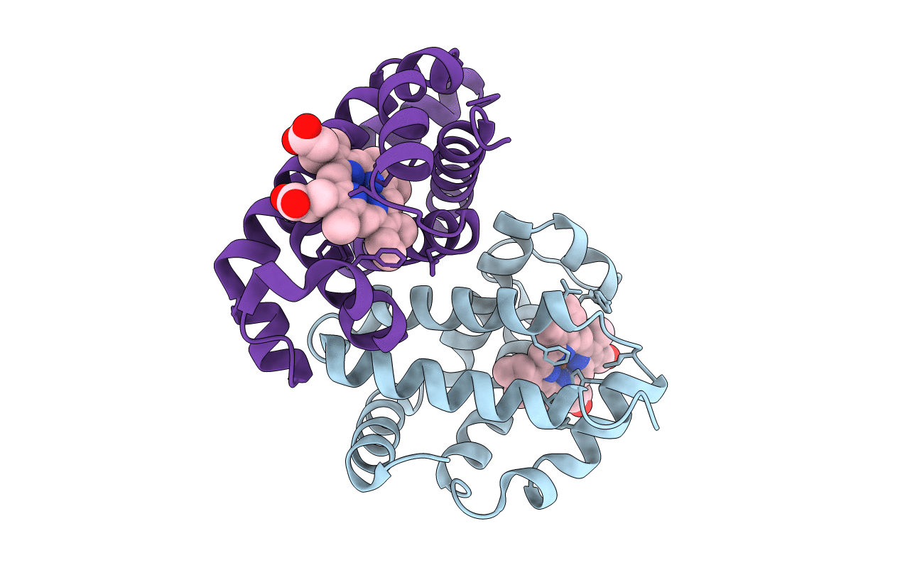
Biological Source:
Source Organism(s):
Peromyscus maniculatus (Taxon ID: 10042)
Expression System(s):
Method Details:
Experimental Method:
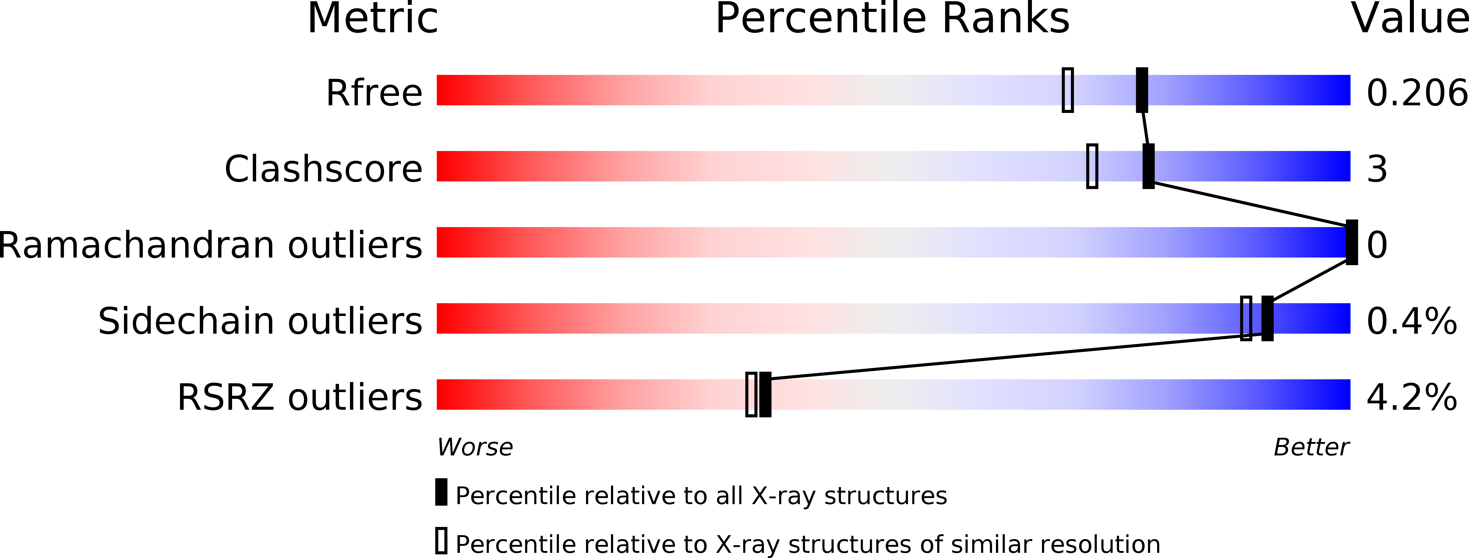
Resolution:
1.78 Å
R-Value Free:
0.20
R-Value Work:
0.16
R-Value Observed:
0.16
Space Group:
C 2 2 21