Deposition Date
2012-09-12
Release Date
2012-11-28
Last Version Date
2024-11-27
Entry Detail
PDB ID:
4H2I
Keywords:
Title:
Human ecto-5'-nucleotidase (CD73): crystal form III (closed) in complex with AMPCP
Biological Source:
Source Organism(s):
Homo sapiens (Taxon ID: 9606)
Expression System(s):
Method Details:
Experimental Method:
Resolution:
2.00 Å
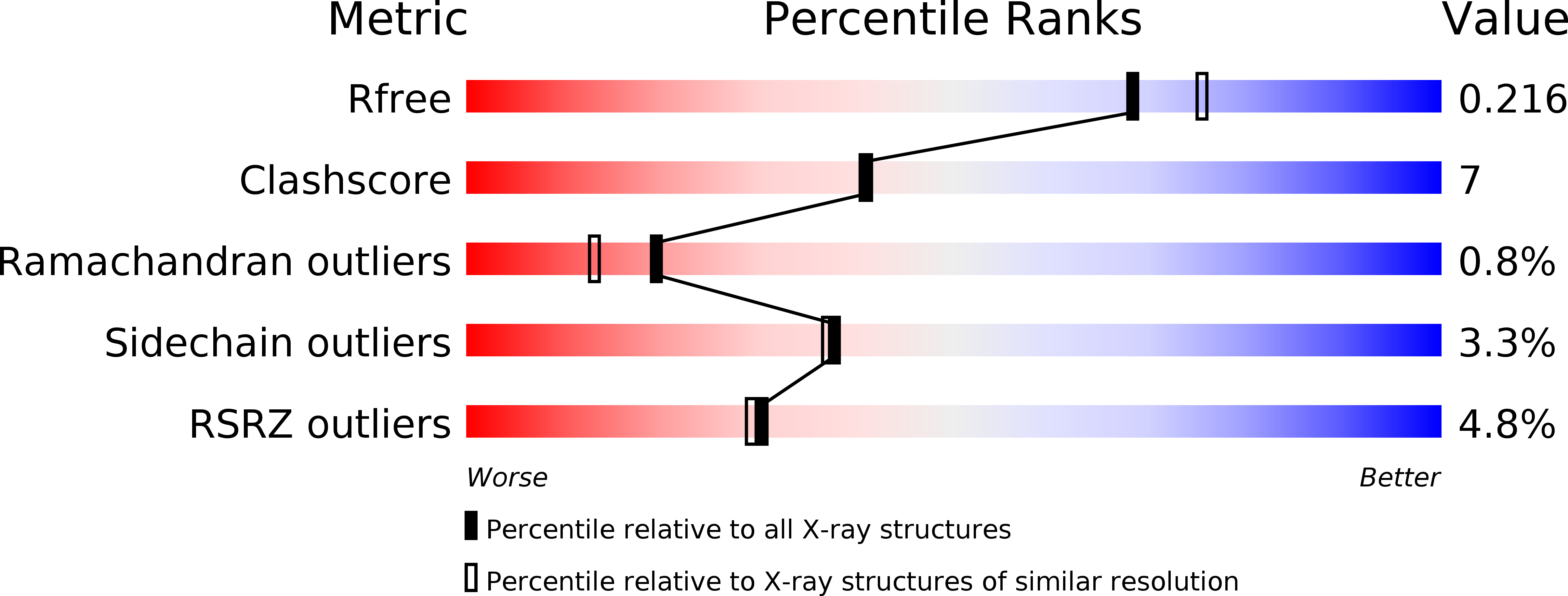
R-Value Free:
0.21
R-Value Work:
0.17
R-Value Observed:
0.17
Space Group:
C 2 2 21