4H0L image
Entry Detail
PDB ID:
4H0L
Keywords:
Title:
Cytochrome b6f Complex Crystal Structure from Mastigocladus laminosus with n-Side Inhibitor NQNO
Biological Source:
Source Organism:
PDB Version:
Deposition Date:
2012-09-08
Release Date:
2013-02-13
Method Details:
Experimental Method:
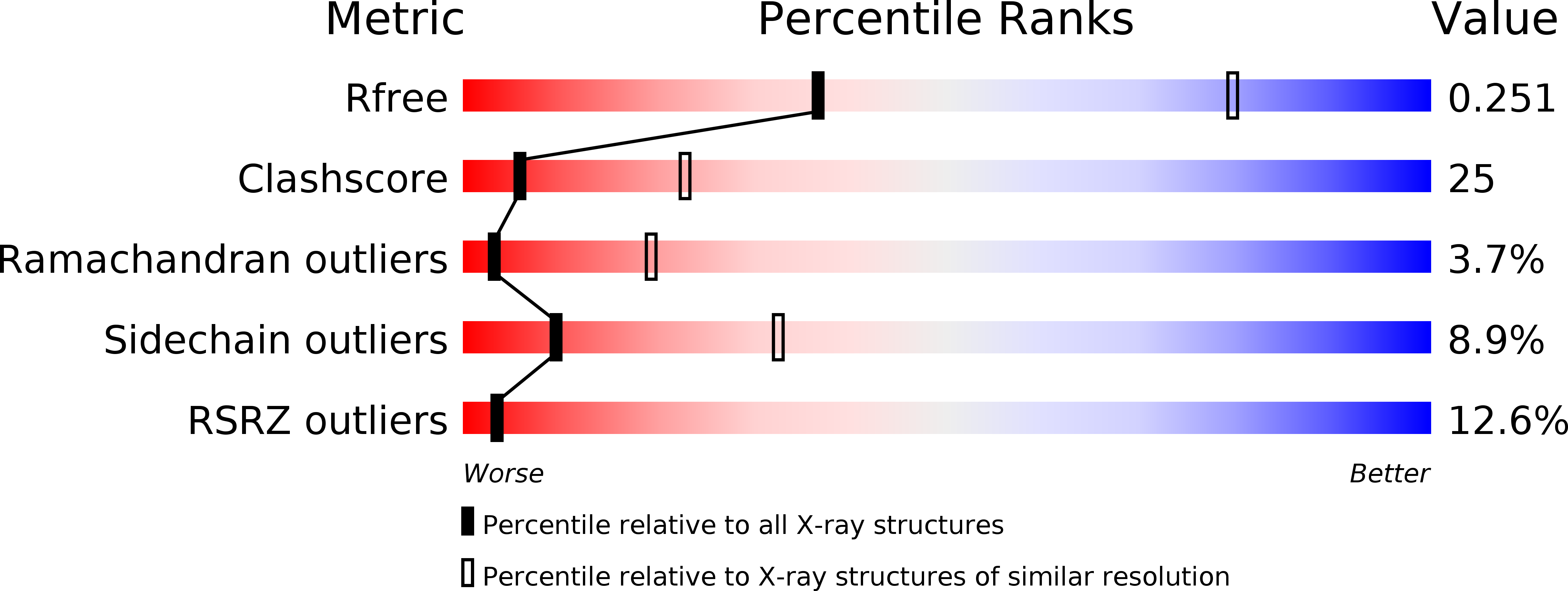
Resolution:
3.25 Å
R-Value Free:
0.24
R-Value Work:
0.21
R-Value Observed:
0.22
Space Group:
P 61 2 2
Macromolecular Entities
Polymer Type:polypeptide(L)
Description:Cytochrome b6
Chain IDs:A
Chain Length:215
Number of Molecules:1
Biological Source:Mastigocladus laminosus
Polymer Type:polypeptide(L)
Description:Cytochrome b6-f complex subunit 4
Chain IDs:B
Chain Length:160
Number of Molecules:1
Biological Source:Mastigocladus laminosus
Polymer Type:polypeptide(L)
Description:Apocytochrome f
Chain IDs:C
Chain Length:289
Number of Molecules:1
Biological Source:Mastigocladus laminosus
Polymer Type:polypeptide(L)
Description:Cytochrome b6-f complex iron-sulfur subunit
Chain IDs:D
Chain Length:179
Number of Molecules:1
Biological Source:Mastigocladus laminosus
Polymer Type:polypeptide(L)
Description:Cytochrome b6-f complex subunit 6
Chain IDs:E
Chain Length:32
Number of Molecules:1
Biological Source:Mastigocladus laminosus
Polymer Type:polypeptide(L)
Description:Cytochrome b6-f complex subunit 7
Chain IDs:F
Chain Length:35
Number of Molecules:1
Biological Source:Mastigocladus laminosus
Polymer Type:polypeptide(L)
Description:Cytochrome b6-f complex subunit 5
Chain IDs:G
Chain Length:37
Number of Molecules:1
Biological Source:Mastigocladus laminosus
Polymer Type:polypeptide(L)
Description:Cytochrome b6-f complex subunit 8
Chain IDs:H
Chain Length:29
Number of Molecules:1
Biological Source:Mastigocladus laminosus
Primary Citation
Quinone-dependent proton transfer pathways in the photosynthetic cytochrome b6f complex
Proc.Natl.Acad.Sci.USA 110 4297 4302 (2013)
PMID: 23440205 DOI: 10.1073/pnas.1222248110

Abstact

As much as two-thirds of the proton gradient used for transmembrane free energy storage in oxygenic photosynthesis is generated by the cytochrome b6f complex. The proton uptake pathway from the electrochemically negative (n) aqueous phase to the n-side quinone binding site of the complex, and a probable route for proton exit to the positive phase resulting from quinol oxidation, are defined in a 2.70-Å crystal structure and in structures with quinone analog inhibitors at 3.07 Å (tridecyl-stigmatellin) and 3.25-Å (2-nonyl-4-hydroxyquinoline N-oxide) resolution. The simplest n-side proton pathway extends from the aqueous phase via Asp20 and Arg207 (cytochrome b6 subunit) to quinone bound axially to heme c(n). On the positive side, the heme-proximal Glu78 (subunit IV), which accepts protons from plastosemiquinone, defines a route for H(+) transfer to the aqueous phase. These pathways provide a structure-based description of the quinone-mediated proton transfer responsible for generation of the transmembrane electrochemical potential gradient in oxygenic photosynthesis.

Legend

Protein

Chemical

Disease

Primary Citation of related structures