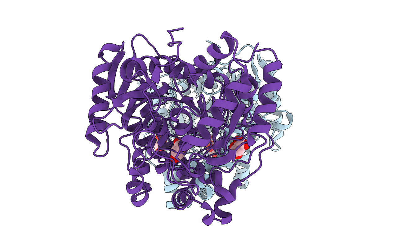
4GYS image
Entry Detail
PDB ID:
4GYS
Keywords:
Title:
Granulibacter bethesdensis allophanate hydrolase co-crystallized with malonate
Biological Source:
Host Organism:
PDB Version:
Deposition Date:
2012-09-05
Release Date:
2013-01-30
Method Details:
Experimental Method:
Resolution:
2.20 Å
R-Value Free:
0.20
R-Value Work:
0.17
R-Value Observed:
0.17
Space Group:
P 61
Macromolecular Entities
Polymer Type:polypeptide(L)
Description:Allophanate hydrolase
Chain IDs:A, B
Chain Length:621
Number of Molecules:2
Biological Source:Granulibacter bethesdensis
Ligand Molecules
Primary Citation
The Structure of Allophanate Hydrolase from Granulibacter bethesdensis Provides Insights into Substrate Specificity in the Amidase Signature Family.
Biochemistry 52 690 700 (2013)
PMID: 23282241 DOI: 10.1021/bi301242m

Abstact

Allophanate hydrolase (AH) catalyzes the hydrolysis of allophanate, an intermediate in atrazine degradation and urea catabolism pathways, to NH(3) and CO(2). AH belongs to the amidase signature family, which is characterized by a conserved block of 130 amino acids rich in Gly and Ser and a Ser-cis-Ser-Lys catalytic triad. In this study, the first structures of AH from Granulibacter bethesdensis were determined, with and without the substrate analogue malonate, to 2.2 and 2.8 Å, respectively. The structures confirm the identity of the catalytic triad residues and reveal an altered dimerization interface that is not conserved in the amidase signature family. The structures also provide insights into previously unrecognized substrate specificity determinants in AH. Two residues, Tyr(299) and Arg(307), are within hydrogen bonding distance of a carboxylate moiety of malonate. Both Tyr(299) and Arg(307) were mutated, and the resulting modified enzymes revealed >3 order of magnitude reductions in both catalytic efficiency and substrate stringency. It is proposed that Tyr(299) and Arg(307) serve to anchor and orient the substrate for attack by the catalytic nucleophile, Ser(172). The structure further suggests the presence of a unique C-terminal domain in AH. While this domain is conserved, it does not contribute to catalysis or to the structural integrity of the core domain, suggesting that it may play a role in mediating transient and specific interactions with the urea carboxylase component of urea amidolyase. Analysis of the AH active site architecture offers new insights into common determinants of catalysis and specificity among divergent members of the amidase signature family.

Legend

Protein

Chemical

Disease

Primary Citation of related structures