Deposition Date
2012-09-05
Release Date
2012-12-19
Last Version Date
2023-09-13
Entry Detail
PDB ID:
4GYJ
Keywords:
Title:
Crystal structure of mutant (D318N) bacillus subtilis family 3 glycoside hydrolase (nagz) in complex with glcnac-murnac (space group P1)
Biological Source:
Source Organism(s):
Bacillus subtilis subsp. subtilis (Taxon ID: 224308)
Expression System(s):
Method Details:
Experimental Method:
Resolution:
1.65 Å
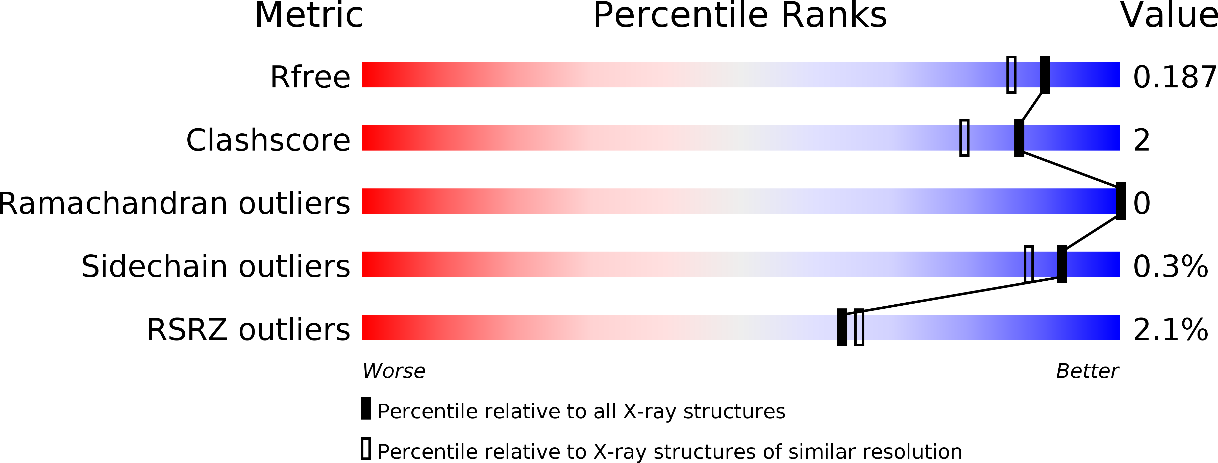
R-Value Free:
0.19
R-Value Work:
0.16
R-Value Observed:
0.16
Space Group:
P 1