4GYE image
Entry Detail
PDB ID:
4GYE
Title:
MDR 769 HIV-1 Protease in Complex with Reduced P1F
Biological Source:
PDB Version:
Deposition Date:
2012-09-05
Release Date:
2013-10-30
Method Details:
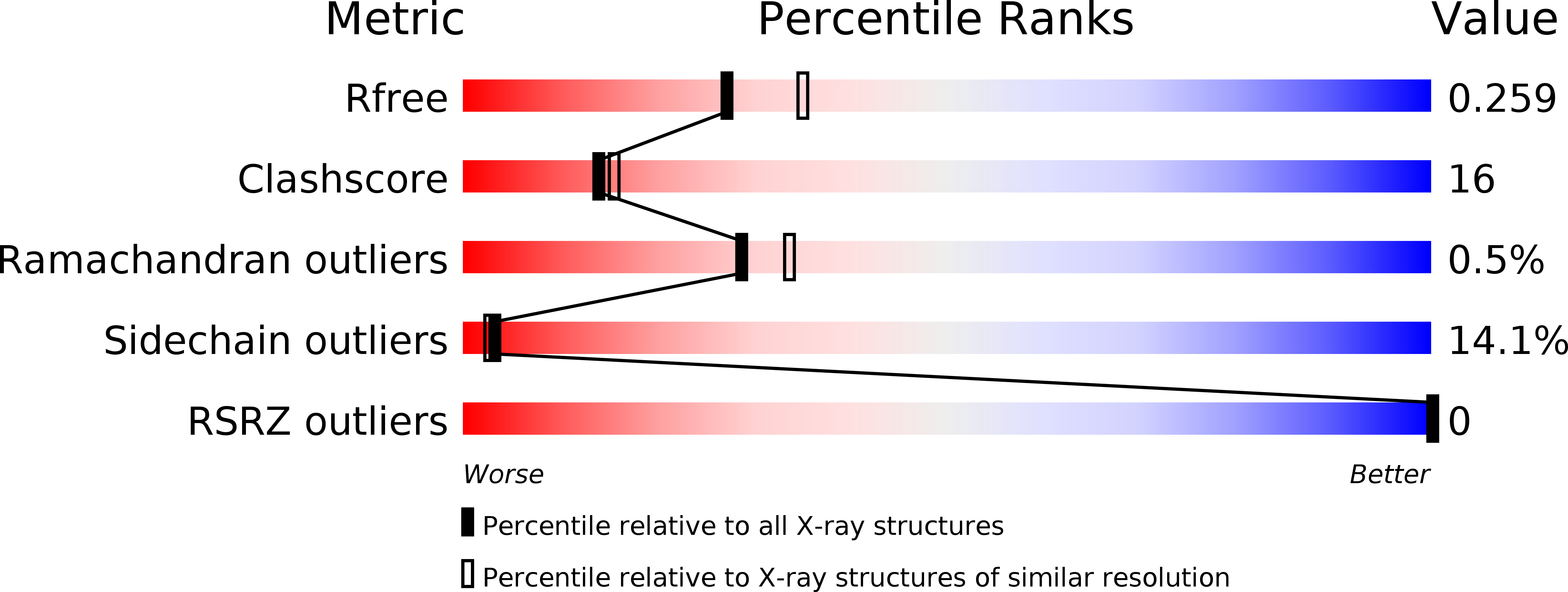
Experimental Method:
Resolution:
2.27 Å
R-Value Free:
0.26
R-Value Work:
0.19
R-Value Observed:
0.19
Space Group:
P 61
Macromolecular Entities
Polymer Type:polypeptide(L)
Description:Protease
Mutations:Q7K, D25N, M36V, A82T, I84V
Chain IDs:A, B
Chain Length:99
Number of Molecules:2
Biological Source:Human immunodeficiency virus 1
Polymer Type:polypeptide(L)
Description:P1F peptide
Chain IDs:C
Chain Length:6
Number of Molecules:1
Biological Source:synthetic construct
Modified Residue
Compound ID Chain ID Parent Comp ID Details 2D Image
NLE C LEU NORLEUCINE
Peptide-like Molecules
PRD_000778
Primary Citation
Ligand modifications to reduce the relative resistance of multi-drug resistant HIV-1 protease.
Bioorg.Med.Chem. 21 7430 7434 (2013)
PMID: 24128815 DOI: 10.1016/j.bmc.2013.09.045

Abstact

Proper proteolytic processing of the HIV-1 Gag/Pol polyprotein is required for HIV infection and viral replication. This feature has made HIV-1 protease an attractive target for antiretroviral drug design for the treatment of HIV-1 infected patients. To examine the role of the P1 and P1'positions of the substrate in inhibitory efficacy of multi-drug resistant HIV-1 protease 769 (MDR 769), we performed a series of structure-function studies. Using the original CA/p2 cleavage site sequence, we generated heptapeptides containing one reduced peptide bond with an L to F and A to F double mutation at P1 and P1' (F-r-F), and an A to F at P1' (L-r-F) resulting in P1/P1' modified ligands. Here, we present an analysis of co-crystal structures of CA/p2 F-r-F, and CA/p2 L-r-F in complex with MDR 769. To examine conformational changes in the complex structure, molecular dynamic (MD) simulations were performed with MDR769-ligand complexes. MD trajectories show the isobutyl group of both the lopinavir analog and the CA/p2 L-r-F substrate cause a conformational change of in the active site of MDR 769. IC50 measurements suggest the non identical P1/P1' ligands (CA/p2 L-r-F and lopinavir analog) are more effective against MDR proteases as opposed to identical P1/P1'ligands. Our results suggest that a non identical P1/P1'composition may be more favorable for the inhibition of MDR 769 as they induce conformational changes in the active site of the enzyme resulting in disruption of the two-fold symmetry of the protease, thus, stabilizing the inhibitor in the active site.

Legend

Protein

Chemical

Disease

Primary Citation of related structures