4GYC image
Deposition Date 2012-09-05
Release Date 2013-05-08
Last Version Date 2024-11-20
Entry Detail
PDB ID:
4GYC
Title:
Structure of the SRII(D75N mutant)/HtrII Complex in I212121 space group ("U" shape)
Biological Source:
Source Organism:
Host Organism:
Method Details:
Experimental Method:
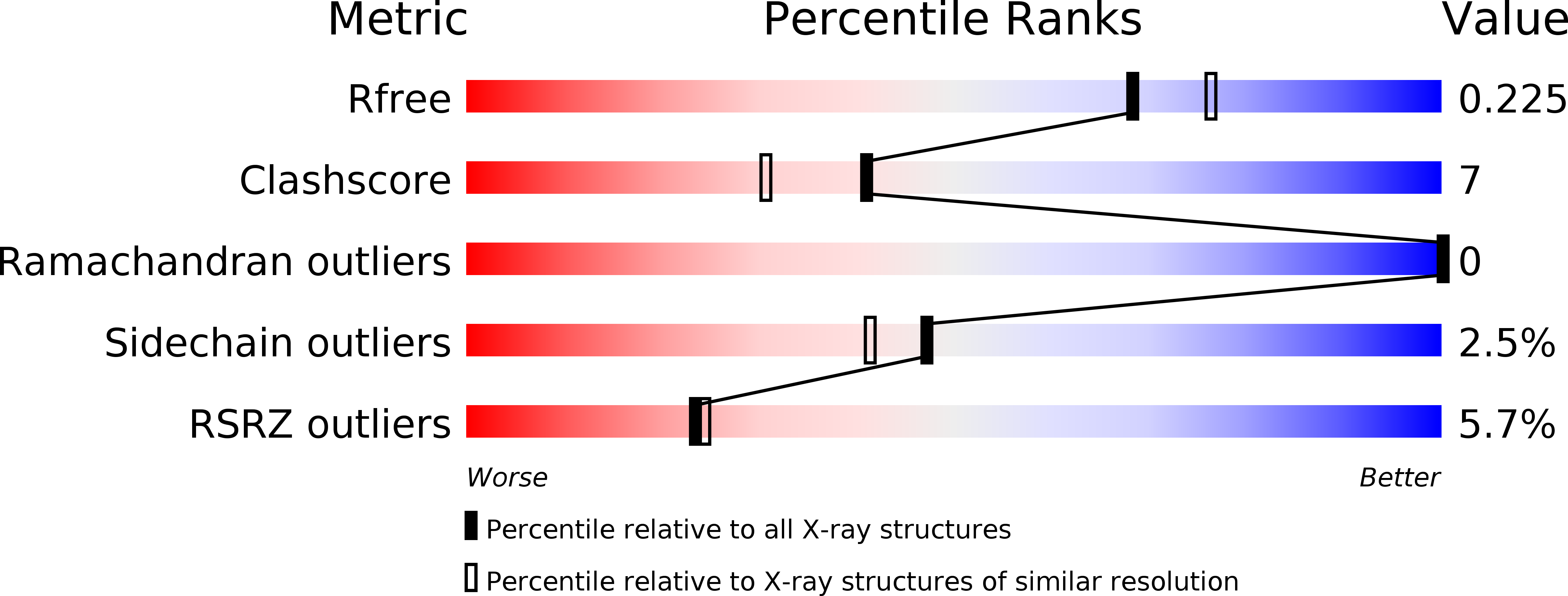
Resolution:
2.05 Å
R-Value Free:
0.22
R-Value Work:
0.20
R-Value Observed:
0.20
Space Group:
I 21 21 21
Macromolecular Entities
Polymer Type:polypeptide(L)
Molecule:Sensory rhodopsin-2
Gene (Uniprot):sop2
Mutations:D75N
Chain IDs:A
Chain Length:245
Number of Molecules:1
Biological Source:Natronomonas pharaonis
Polymer Type:polypeptide(L)
Molecule:Sensory rhodopsin II transducer
Gene (Uniprot):htr2
Chain IDs:B
Chain Length:135
Number of Molecules:1
Biological Source:Natronomonas pharaonis
Primary Citation
Ground state structure of D75N mutant of sensory rhodopsin II in complex with its cognate transducer.
J Photochem Photobiol B 123C 55 58 (2013)
PMID: 23619282 DOI: 10.1016/j.jphotobiol.2013.03.008

Abstact

The complex of sensory rhodopsin II (NpSRII) with its cognate transducer (NpHtrII) mediates negative phototaxis in halobacteria Natronomonas pharaonis. Upon light activation NpSRII triggers, by means of NpHtrII, a signal transduction chain homologous to the two component system in eubacterial chemotaxis. Here we report on the crystal structure of the ground state of the mutant NpSRII-D75N/NpHtrII complex in the space group I212121. Mutations of this aspartic acid in light-driven proton pumps dramatically modify or/and inhibit protein functions. However, in vivo studies show that the similar D75N mutation retains functionality of the NpSRII/NpHtrII complex. The structure provides the molecular basis for the explanation of the unexpected observation that the wild and the mutant complexes display identical physiological response on light excitation.

Legend

Protein

Chemical

Disease

Primary Citation of related structures