
Deposition Date
2012-09-04
Release Date
2013-04-03
Last Version Date
2023-11-08
Entry Detail
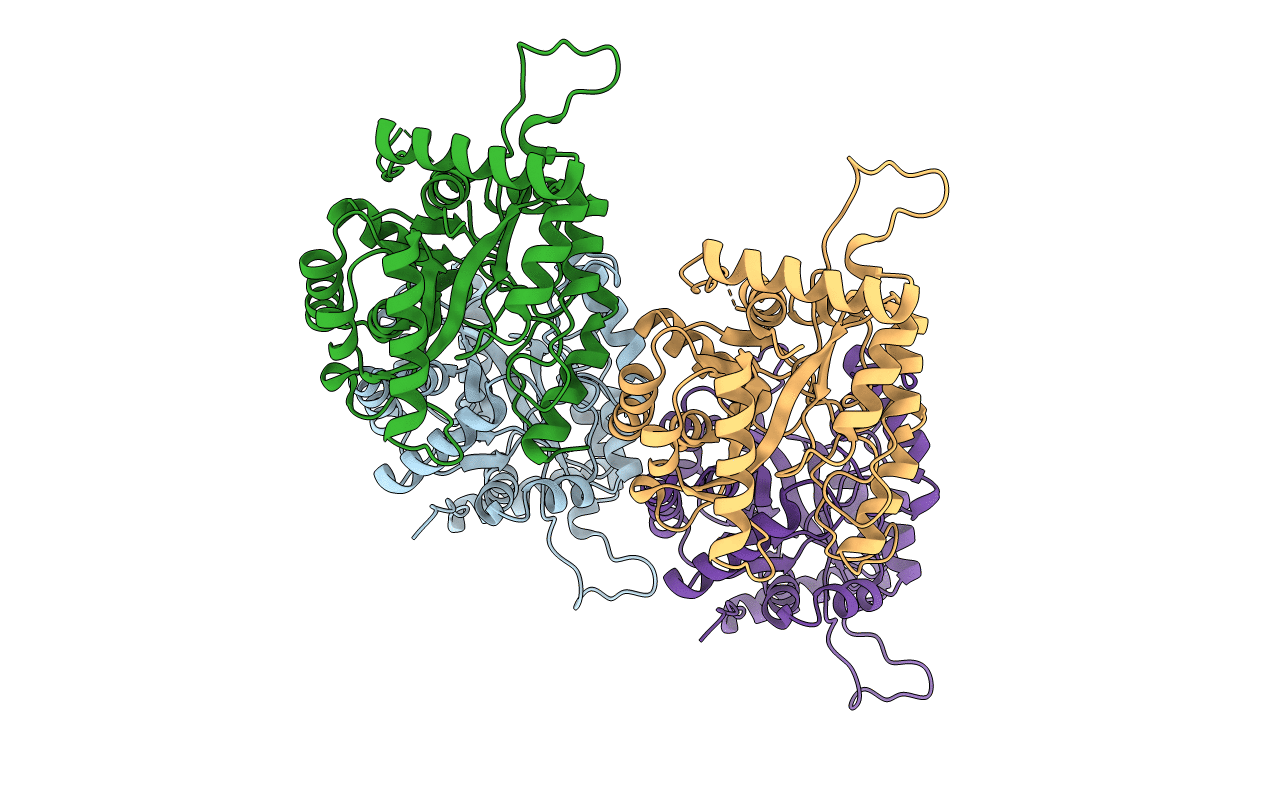
PDB ID:
4GX9
Keywords:
Title:
Crystal structure of a DNA polymerase III alpha-epsilon chimera
Biological Source:
Source Organism(s):
Escherichia coli (Taxon ID: 83333)
Expression System(s):
Method Details:
Experimental Method:
Resolution:
2.15 Å
R-Value Free:
0.29
R-Value Work:
0.22
R-Value Observed:
0.22
Space Group:
P 1 21 1
