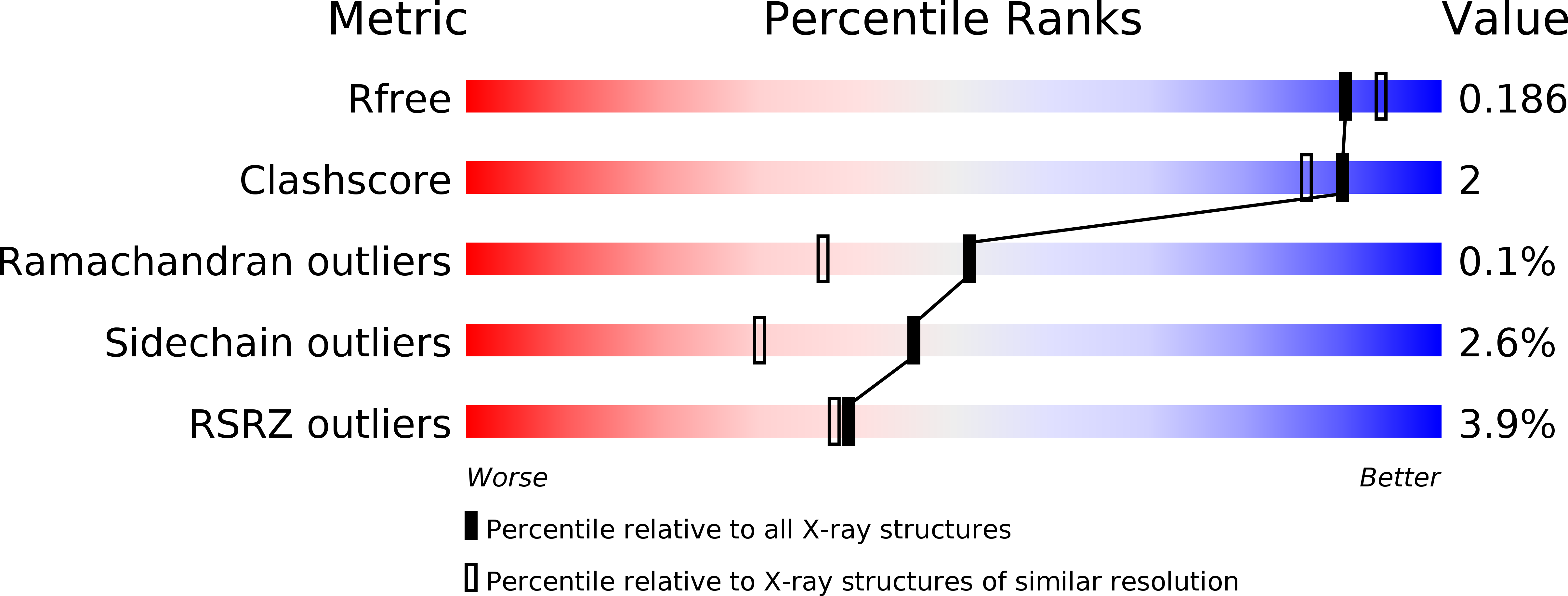
4GWM image
Entry Detail
PDB ID:
4GWM
Keywords:
Title:
Crystal structure of human promeprin beta
Biological Source:
Source Organism:
Host Organism:
PDB Version:
Deposition Date:
2012-09-03
Release Date:
2012-09-19
Method Details:
Experimental Method:
Resolution:
1.85 Å
R-Value Free:
0.18
R-Value Work:
0.16
R-Value Observed:
0.16
Space Group:
P 1
Macromolecular Entities
Polymer Type:polypeptide(L)
Description:Meprin A subunit beta
Mutations:T23P, P24W
Chain IDs:A, B
Chain Length:592
Number of Molecules:2
Biological Source:Homo sapiens
Modified Residue
Compound ID Chain ID Parent Comp ID Details 2D Image
ASN B ASN GLYCOSYLATION SITE
Primary Citation
Structural basis for the sheddase function of human meprin beta metalloproteinase at the plasma membrane
Proc.Natl.Acad.Sci.USA 109 16131 16136 (2012)
PMID: 22988105 DOI: 10.1073/pnas.1211076109

Abstact

Ectodomain shedding at the cell surface is a major mechanism to regulate the extracellular and circulatory concentration or the activities of signaling proteins at the plasma membrane. Human meprin β is a 145-kDa disulfide-linked homodimeric multidomain type-I membrane metallopeptidase that sheds membrane-bound cytokines and growth factors, thereby contributing to inflammatory diseases, angiogenesis, and tumor progression. In addition, it cleaves amyloid precursor protein (APP) at the β-secretase site, giving rise to amyloidogenic peptides. We have solved the X-ray crystal structure of a major fragment of the meprin β ectoprotein, the first of a multidomain oligomeric transmembrane sheddase, and of its zymogen. The meprin β dimer displays a compact shape, whose catalytic domain undergoes major rearrangement upon activation, and reveals an exosite and a sugar-rich channel, both of which possibly engage in substrate binding. A plausible structure-derived working mechanism suggests that substrates such as APP are shed close to the plasma membrane surface following an "N-like" chain trace.

Legend

Protein

Chemical

Disease

Primary Citation of related structures