Deposition Date
2012-09-01
Release Date
2012-09-26
Last Version Date
2024-11-27
Entry Detail
PDB ID:
4GW9
Keywords:
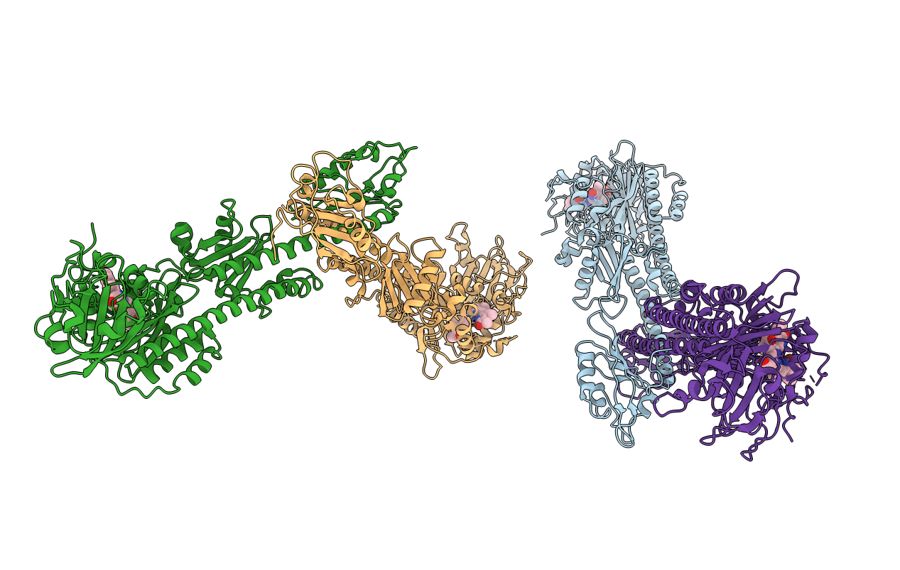
Title:
Structure of a bacteriophytochrome and light-stimulated protomer swapping with a gene repressor
Biological Source:
Source Organism:
Rhodopseudomonas palustris (Taxon ID: 258594)
Host Organism:
Method Details:
Experimental Method:
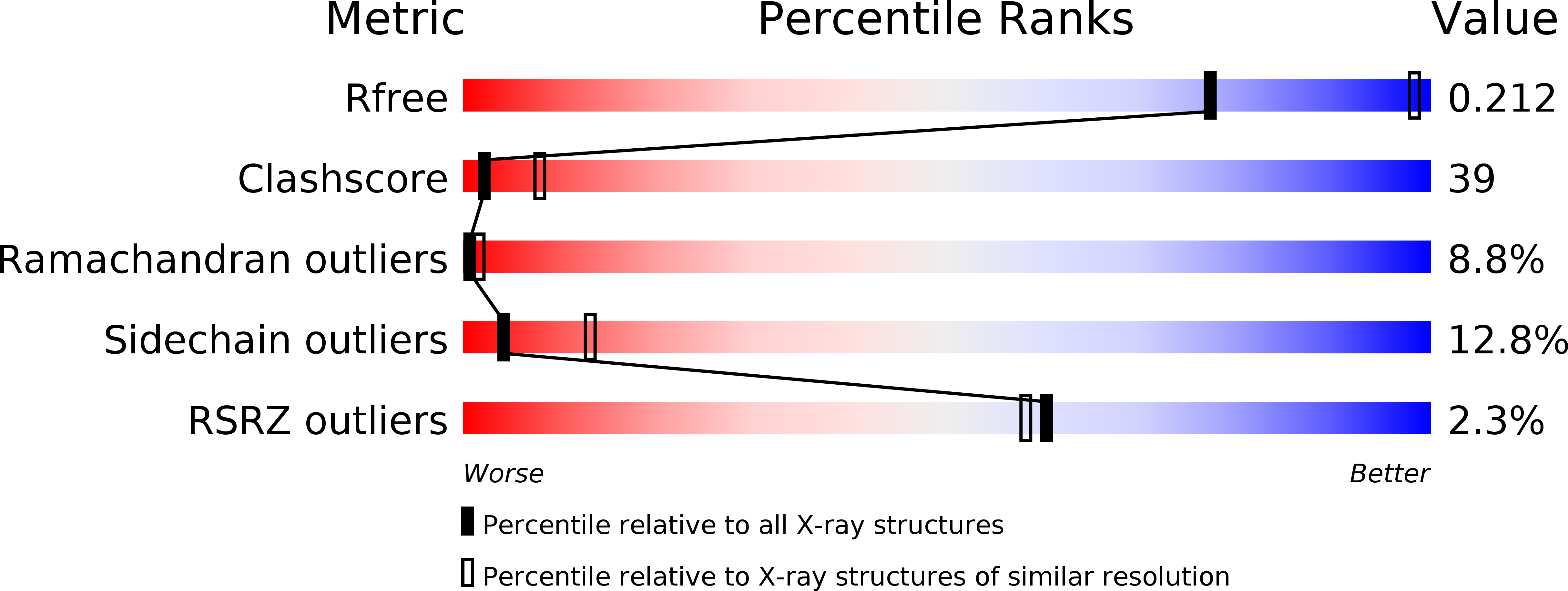
Resolution:
2.90 Å
R-Value Free:
0.24
R-Value Work:
0.19
R-Value Observed:
0.20
Space Group:
P 1 21 1