4GSW image
Deposition Date 2012-08-28
Release Date 2012-12-12
Last Version Date 2023-09-13
Entry Detail
PDB ID:
4GSW
Keywords:
Title:
Crystal structure of ubiquitin from Entamoeba histolytica to 2.15 Angstrom
Biological Source:
Source Organism:
Host Organism:
Method Details:
Experimental Method:
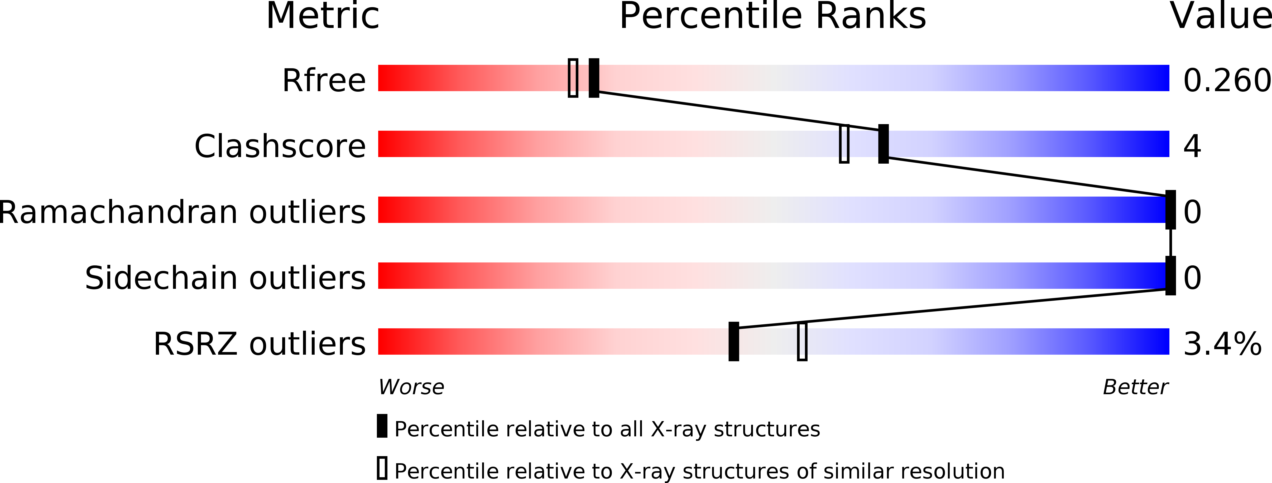
Resolution:
2.15 Å
R-Value Free:
0.25
R-Value Work:
0.19
R-Value Observed:
0.19
Space Group:
P 21 21 21
Macromolecular Entities
Structures with similar UniProt ID
Protein Blast
Polymer Type:polypeptide(L)
Molecule:Ubiquitin
Gene (Uniprot):EHI_083270, EHI_083410, EHI_156660, EHI_178340
Chain IDs:A, B
Chain Length:80
Number of Molecules:2
Biological Source:Entamoeba histolytica
Ligand Molecules
Primary Citation
Structural Determinants of Ubiquitin Conjugation in Entamoeba histolytica.
J.Biol.Chem. 288 2290 2302 (2013)
PMID: 23209297 DOI: 10.1074/jbc.M112.417337

Abstact

Ubiquitination is important for numerous cellular processes in most eukaryotic organisms, including cellular proliferation, development, and protein turnover by the proteasome. The intestinal parasite Entamoeba histolytica harbors an extensive ubiquitin-proteasome system. Proteasome inhibitors are known to impair parasite proliferation and encystation, suggesting the ubiquitin-proteasome pathway as a viable therapeutic target. However, no functional studies of the E. histolytica ubiquitination enzymes have yet emerged. Here, we have cloned and characterized multiple E. histolytica ubiquitination components, spanning ubiquitin and its activating (E1), conjugating (E2), and ligating (E3) enzymes. Crystal structures of EhUbiquitin reveal a clustering of unique residues on the α1 helix surface, including an eighth surface lysine not found in other organisms, which may allow for a unique polyubiquitin linkage in E. histolytica. EhUbiquitin is activated by and forms a thioester bond with EhUba1 (E1) in vitro, in an ATP- and magnesium-dependent fashion. EhUba1 exhibits a greater maximal initial velocity of pyrophosphate:ATP exchange than its human homolog, suggesting different kinetics of ubiquitin activation in E. histolytica. EhUba1 engages the E2 enzyme EhUbc5 through its ubiquitin-fold domain to transfer the EhUbiquitin thioester. However, EhUbc5 has a >10-fold preference for EhUba1∼Ub compared with unconjugated EhUba1. A crystal structure of EhUbc5 allowed prediction of a noncovalent "backside" interaction with EhUbiquitin and E3 enzymes. EhUbc5 selectively engages EhRING1 (E3) to the exclusion of two HECT family E3 ligases, and mutagenesis indicates a conserved mode of E2/RING-E3 interaction in E. histolytica.

Legend

Protein

Chemical

Disease

Primary Citation of related structures