4GQC image
Entry Detail
PDB ID:
4GQC
Keywords:
Title:
Crystal Structure of Aeropyrum pernix Peroxiredoxin Q Enzyme in Fully-Folded and Locally-Unfolded Conformations
Biological Source:
Source Organism:
Host Organism:
PDB Version:
Deposition Date:
2012-08-22
Release Date:
2012-10-24
Method Details:
Experimental Method:
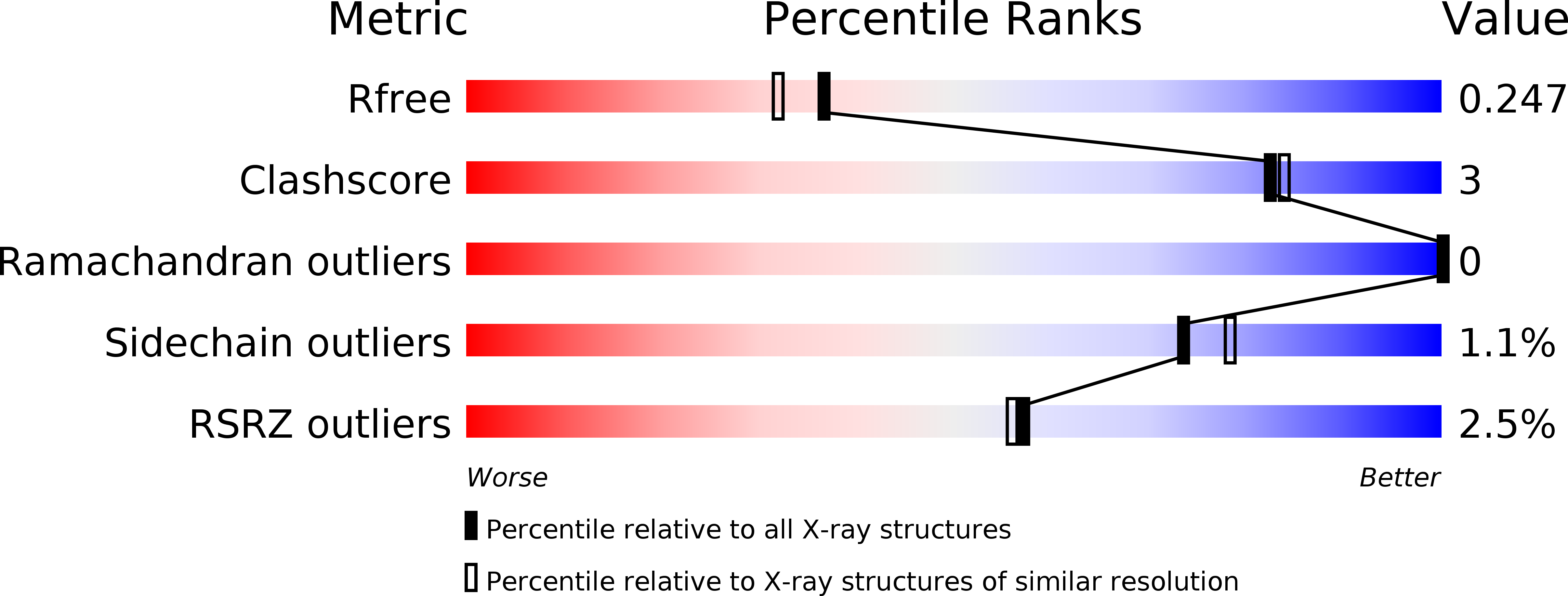
Resolution:
2.00 Å
R-Value Free:
0.23
R-Value Work:
0.19
R-Value Observed:
0.19
Space Group:
P 41 2 2
Macromolecular Entities
Polymer Type:polypeptide(L)
Description:Thiol peroxidase
Chain IDs:A, B, C, D
Chain Length:164
Number of Molecules:4
Biological Source:Aeropyrum pernix
Primary Citation
Mapping the Active Site Helix-to-Strand Conversion of CxxxxC Peroxiredoxin Q Enzymes.
Biochemistry 51 7638 7650 (2012)
PMID: 22928725 DOI: 10.1021/bi301017s

Abstact

Peroxiredoxins (Prx) make up a family of enzymes that reduce peroxides using a peroxidatic cysteine residue; among these, members of the PrxQ subfamily are proposed to be the most ancestral-like yet are among the least characterized. In many PrxQ enzymes, a second "resolving" cysteine is located five residues downstream from the peroxidatic Cys, and these residues form a disulfide during the catalytic cycle. Here, we describe three hyperthermophilic PrxQ crystal structures originally determined by the RIKEN structural genomics group. We reprocessed the diffraction data and conducted further refinement to yield models with R(free) values lowered by 2.3-7.2% and resolution extended by 0.2-0.3 Å, making one, at 1.4 Å, one of the best resolved peroxiredoxins to date. Comparisons of two matched thiol and disulfide forms reveal that the active site conformational change required for disulfide formation involves a transition of ~20 residues from a pair of α-helices to a β-hairpin and 3(10)-helix. Each conformation has ~10 residues with a high level of disorder providing slack that allows the dramatic shift, and the two conformations are anchored to the protein core by distinct nonpolar side chains that fill three hydrophobic pockets. Sequence conservation patterns confirm the importance of these and a few additional residues for function. From a broader perspective, this study raises the provocative question of how to make use of the valuable information in the Protein Data Bank generated by structural genomics projects but not described in the literature, perhaps remaining unrecognized and certainly underutilized.

Legend

Protein

Chemical

Disease

Primary Citation of related structures