Deposition Date
2012-08-21
Release Date
2013-02-27
Last Version Date
2024-11-20
Entry Detail
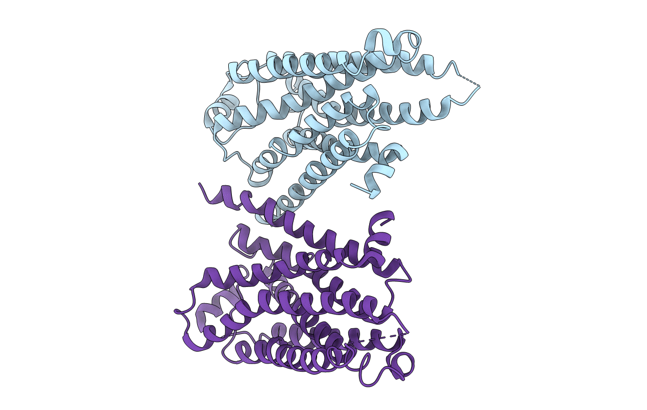
Biological Source:
Source Organism(s):
Meleagris gallopavo (Taxon ID: 9103)
Expression System(s):
Method Details:
Experimental Method:
Resolution:
3.50 Å
R-Value Free:
0.35
R-Value Work:
0.30
R-Value Observed:
0.31
Space Group:
C 1 2 1