4GPN image
Deposition Date 2012-08-21
Release Date 2012-10-03
Last Version Date 2023-09-13
Entry Detail
PDB ID:
4GPN
Keywords:
Title:
The crystal structure of 6-P-beta-D-Glucosidase (E375Q mutant) from Streptococcus mutans UA150 in complex with Gentiobiose 6-phosphate.
Biological Source:
Source Organism:
Host Organism:
Method Details:
Experimental Method:
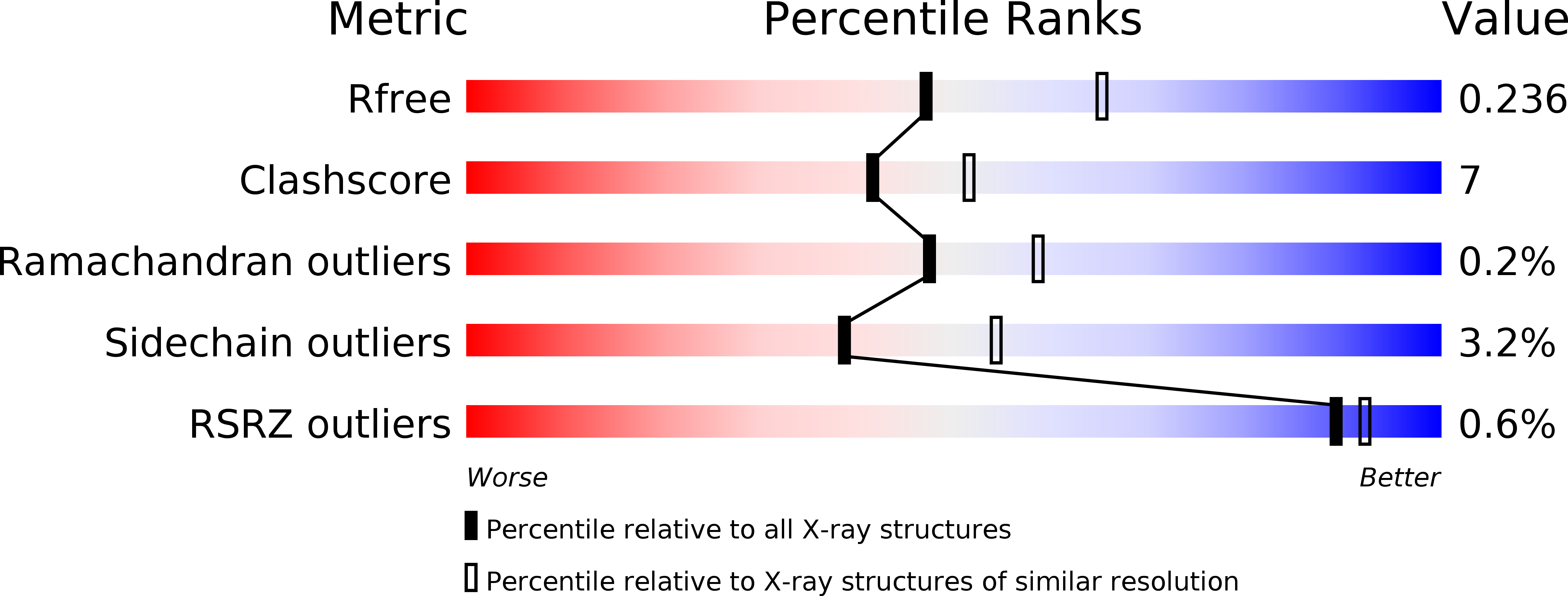
Resolution:
2.29 Å
R-Value Free:
0.23
R-Value Work:
0.17
R-Value Observed:
0.18
Space Group:
P 1 21 1
Macromolecular Entities
Structures with similar UniProt ID
Protein Blast
Polymer Type:polypeptide(L)
Molecule:6-phospho-beta-D-Glucosidase
Gene (Uniprot):bgl
Mutagens:E375Q
Chain IDs:A, B
Chain Length:480
Number of Molecules:2
Biological Source:Streptococcus mutans
Peptide-like Molecules
PRD_900069
Primary Citation
GH1-family 6-P-beta-glucosidases from human microbiome lactic acid bacteria.
Acta Crystallogr. D Biol. Crystallogr. 69 451 463 (2013)
PMID: 23519420 DOI: 10.1107/S0907444912049608

Abstact

In lactic acid bacteria and other bacteria, carbohydrate uptake is mostly governed by phosphoenolpyruvate-dependent phosphotransferase systems (PTSs). PTS-dependent translocation through the cell membrane is coupled with phosphorylation of the incoming sugar. After translocation through the bacterial membrane, the β-glycosidic bond in 6'-P-β-glucoside is cleaved, releasing 6-P-β-glucose and the respective aglycon. This reaction is catalyzed by 6-P-β-glucosidases, which belong to two glycoside hydrolase (GH) families: GH1 and GH4. Here, the high-resolution crystal structures of GH1 6-P-β-glucosidases from Lactobacillus plantarum (LpPbg1) and Streptococcus mutans (SmBgl) and their complexes with ligands are reported. Both enzymes show hydrolytic activity towards 6'-P-β-glucosides. The LpPbg1 structure has been determined in an apo form as well as in a complex with phosphate and a glucose molecule corresponding to the aglycon molecule. The S. mutans homolog contains a sulfate ion in the phosphate-dedicated subcavity. SmBgl was also crystallized in the presence of the reaction product 6-P-β-glucose. For a mutated variant of the S. mutans enzyme (E375Q), the structure of a 6'-P-salicin complex has also been determined. The presence of natural ligands enabled the definition of the structural elements that are responsible for substrate recognition during catalysis.

Legend

Protein

Chemical

Disease

Primary Citation of related structures
Feedback Form
Name
Email
Institute
Feedback