4GP0 image
Deposition Date 2012-08-20
Release Date 2012-11-28
Last Version Date 2023-09-13
Entry Detail
PDB ID:
4GP0
Keywords:
Title:
The crystal structure of human fascin 1 R149A K150A R151A mutant
Biological Source:
Source Organism:
Homo sapiens (Taxon ID: 9606)
Host Organism:
Method Details:
Experimental Method:
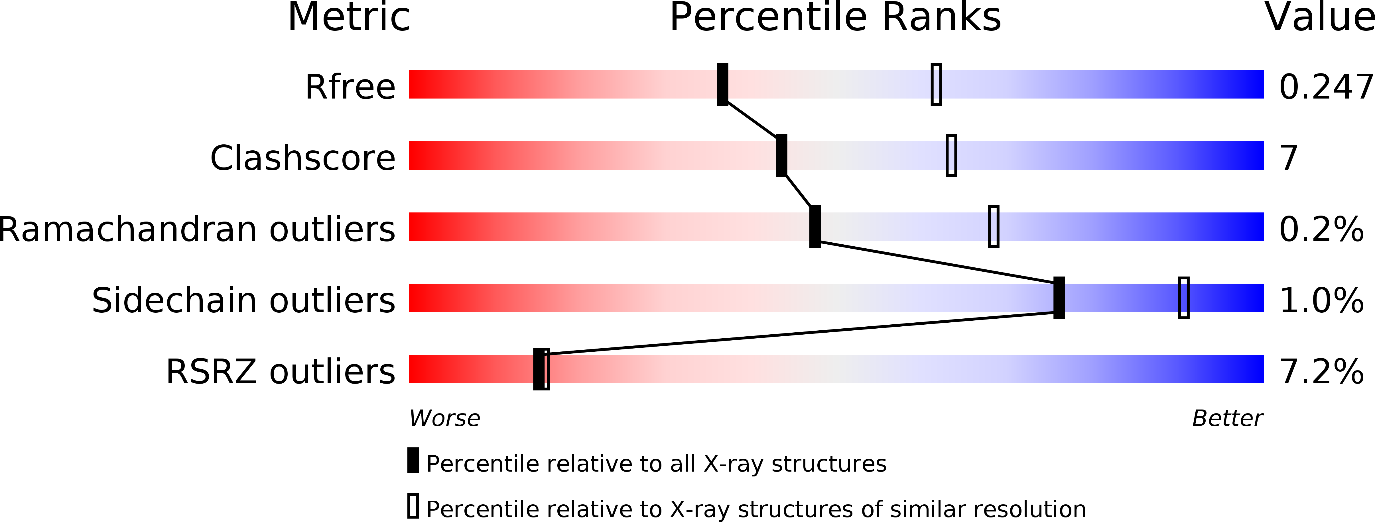
Resolution:
2.50 Å
R-Value Free:
0.24
R-Value Work:
0.20
R-Value Observed:
0.20
Space Group:
C 1 2 1
Macromolecular Entities
Polymer Type:polypeptide(L)
Molecule:Fascin
Gene (Uniprot):FSCN1
Mutations:R149A K150A R151A
Chain IDs:A, B
Chain Length:493
Number of Molecules:2
Biological Source:Homo sapiens
Primary Citation
Molecular mechanism of fascin function in filopodial formation.
J.Biol.Chem. 288 274 284 (2013)
PMID: 23184945 DOI: 10.1074/jbc.M112.427971

Abstact

Filopodia are cell surface protrusions that are essential for cell migration. This finger-like structure is supported by rigid tightly bundled actin filaments. The protein responsible for actin bundling in filopodia is fascin. However, the mechanism by which fascin functions in filopodial formation is not clear. Here we provide biochemical, cryo-electron tomographic, and x-ray crystal structural data demonstrating the unique structural characteristics of fascin. Systematic mutagenesis studies on 100 mutants of fascin indicate that there are two major actin-binding sites on fascin. Crystal structures of four fascin mutants reveal concerted conformational changes in fascin from inactive to active states in the process of actin bundling. Mutations in any one of the actin-binding sites impair the cellular function of fascin in filopodial formation. Altogether, our data reveal the molecular mechanism of fascin function in filopodial formation.

Legend

Protein

Chemical

Disease

Primary Citation of related structures