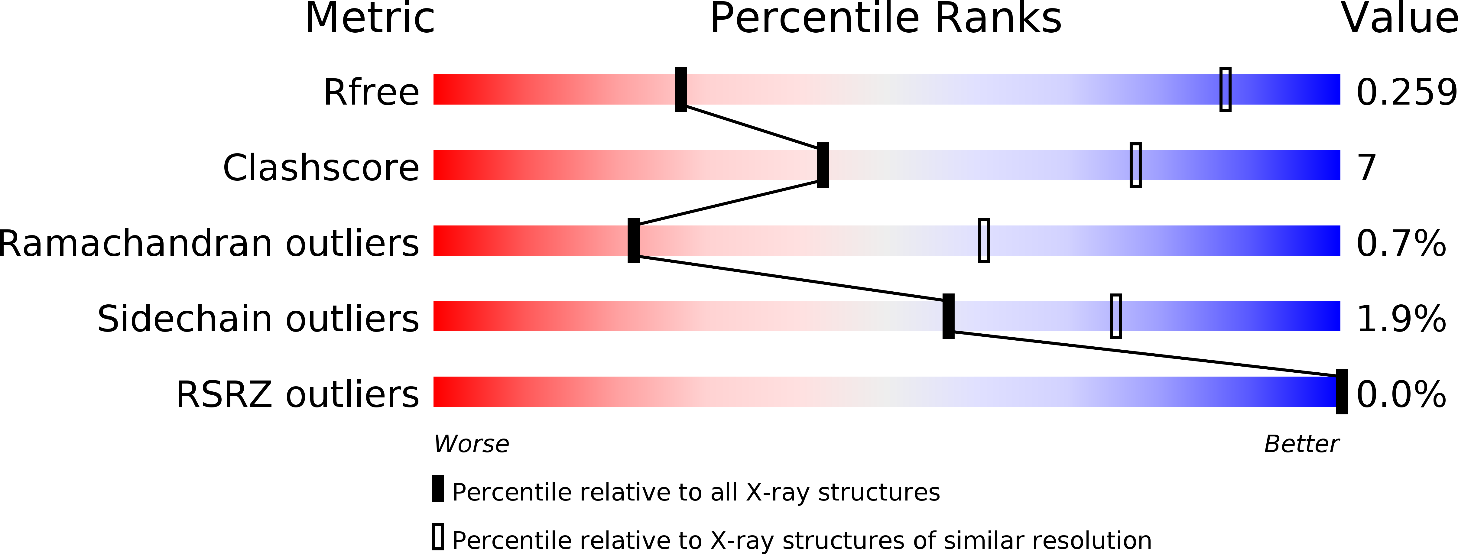
4GNK image
Deposition Date 2012-08-17
Release Date 2013-02-06
Last Version Date 2023-09-13
Entry Detail
PDB ID:
4GNK
Title:
Crystal structure of Galphaq in complex with full-length human PLCbeta3
Biological Source:
Source Organism:
Mus musculus (Taxon ID: 10090)
Homo sapiens (Taxon ID: 9606)
Host Organism:
Method Details:
Experimental Method:
Resolution:
4.00 Å
R-Value Free:
0.25
R-Value Work:
0.21
R-Value Observed:
0.21
Space Group:
P 21 21 21
Macromolecular Entities
Structures with similar UniProt ID
Protein Blast
Polymer Type:polypeptide(L)
Molecule:Guanine nucleotide-binding protein G(q) subunit alpha
Gene (Uniprot):Gnaq
Chain IDs:A, C
Chain Length:353
Number of Molecules:2
Biological Source:Mus musculus
Structures with similar UniProt ID
Protein Blast
Polymer Type:polypeptide(L)
Molecule:1-phosphatidylinositol 4,5-bisphosphate phosphodiesterase beta-3
Gene (Uniprot):PLCB3
Chain IDs:B, D
Chain Length:1235
Number of Molecules:2
Biological Source:Homo sapiens
Structures with similar UniProt ID
Protein Blast
Polymer Type:polypeptide(L)
Molecule:1-phosphatidylinositol 4,5-bisphosphate phosphodiesterase beta-3
Gene (Uniprot):PLCB3
Chain IDs:E
Chain Length:259
Number of Molecules:1
Biological Source:Homo sapiens
Primary Citation
Full-length G alpha (q)-phospholipase C-beta 3 structure reveals interfaces of the C-terminal coiled-coil domain.
Nat.Struct.Mol.Biol. 20 355 362 (2013)
PMID: 23377541 DOI: 10.1038/nsmb.2497

Abstact

Phospholipase C-β (PLCβ) is directly activated by Gαq, but the molecular basis for how its distal C-terminal domain (CTD) contributes to maximal activity is poorly understood. Herein we present both the crystal structure and cryo-EM three-dimensional reconstructions of human full-length PLCβ3 in complex with mouse Gαq. The distal CTD forms an extended monomeric helical bundle consisting of three antiparallel segments with structural similarity to membrane-binding bin-amphiphysin-Rvs (BAR) domains. Sequence conservation of the distal CTD suggests putative membrane and protein interaction sites, the latter of which bind the N-terminal helix of Gαq in both the crystal structure and cryo-EM reconstructions. Functional analysis suggests that the distal CTD has roles in membrane targeting and in optimizing the orientation of the catalytic core at the membrane for maximal rates of lipid hydrolysis.

Legend

Protein

Chemical

Disease

Primary Citation of related structures