Deposition Date
2012-08-13
Release Date
2012-10-03
Last Version Date
2024-02-28
Entry Detail
PDB ID:
4GKQ
Keywords:
Title:
Structure of the neck and C-terminal motor homology domain of ViK1 from Candida glabrata
Biological Source:
Source Organism(s):
Candida glabrata (Taxon ID: 284593)
Expression System(s):
Method Details:
Experimental Method:
Resolution:
2.99 Å
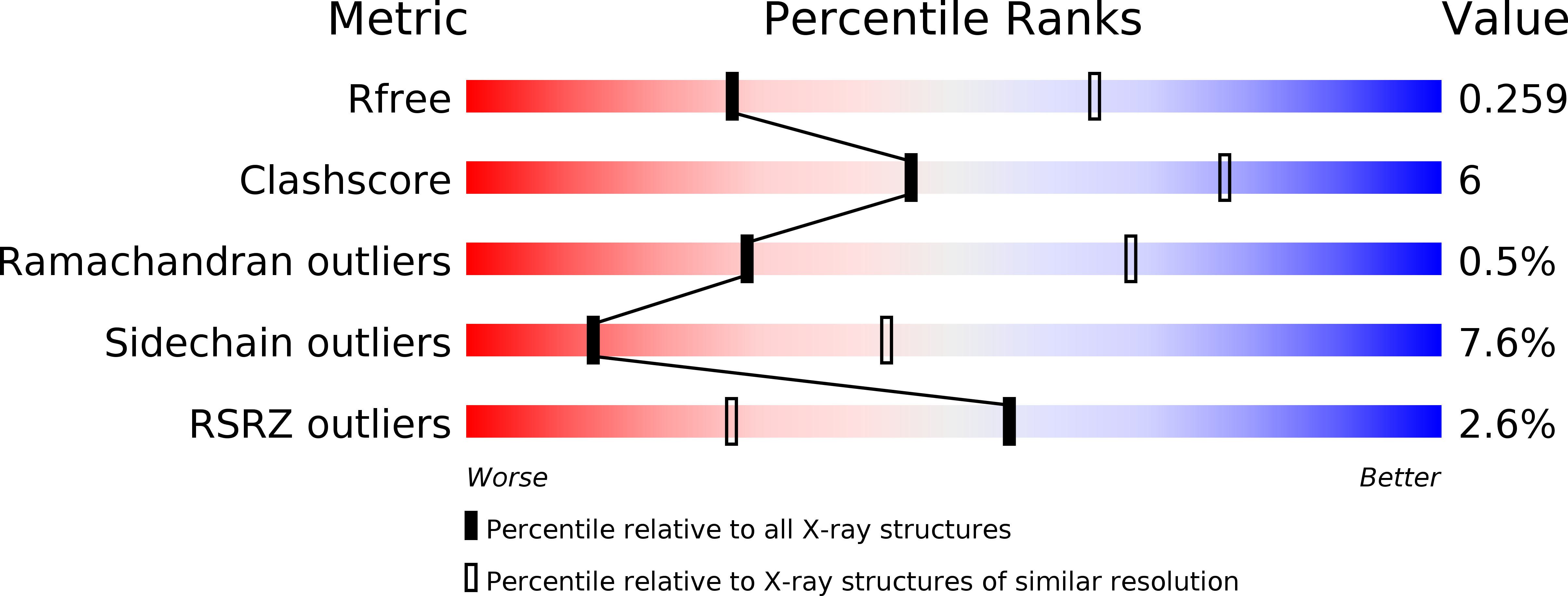
R-Value Free:
0.25
R-Value Work:
0.21
R-Value Observed:
0.21
Space Group:
H 3