Deposition Date
2012-08-09
Release Date
2013-08-14
Last Version Date
2023-09-13
Entry Detail
PDB ID:
4GJ4
Keywords:
Title:
The Crystal Structure of the soluble Guanylate Cyclase PAS alpha domain from Manduca sexta
Biological Source:
Source Organism(s):
Manduca sexta (Taxon ID: 7130)
Expression System(s):
Method Details:
Experimental Method:
Resolution:
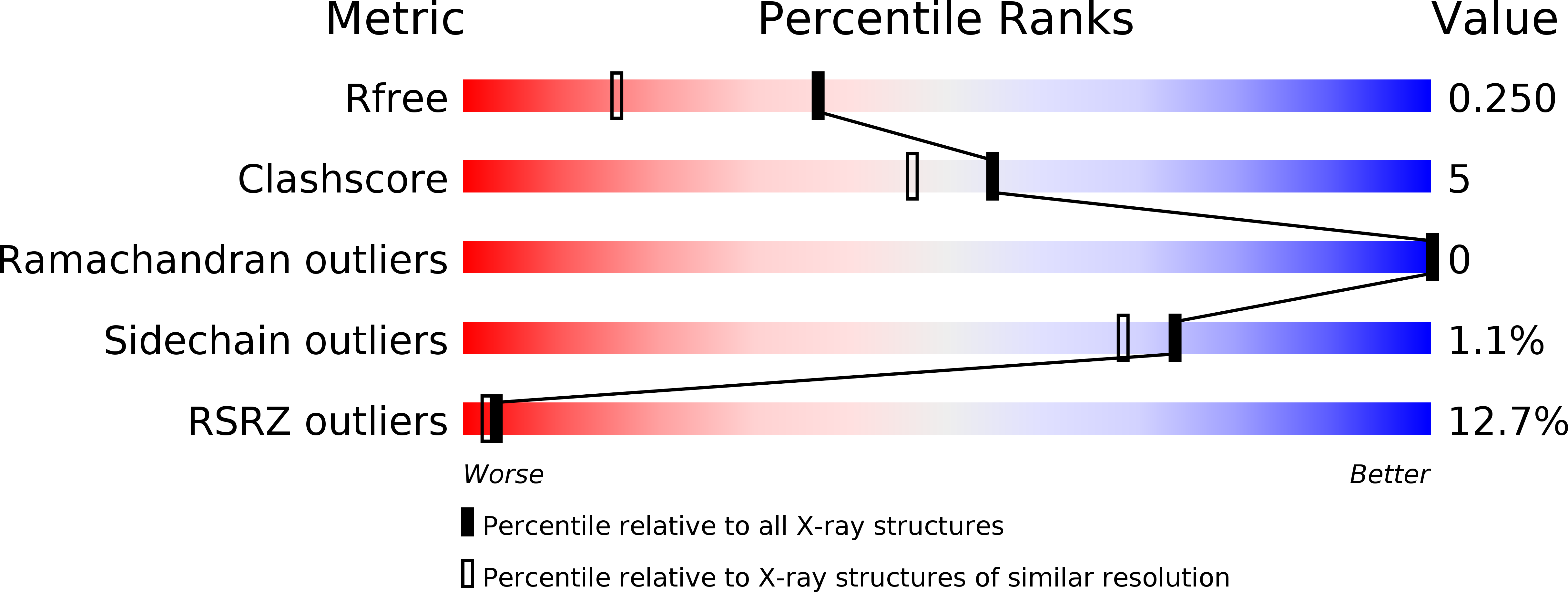
1.80 Å
R-Value Free:
0.24
R-Value Work:
0.19
R-Value Observed:
0.19
Space Group:
H 3 2