Deposition Date
2012-08-08
Release Date
2012-09-19
Last Version Date
2024-10-16
Entry Detail
PDB ID:
4GIP
Keywords:
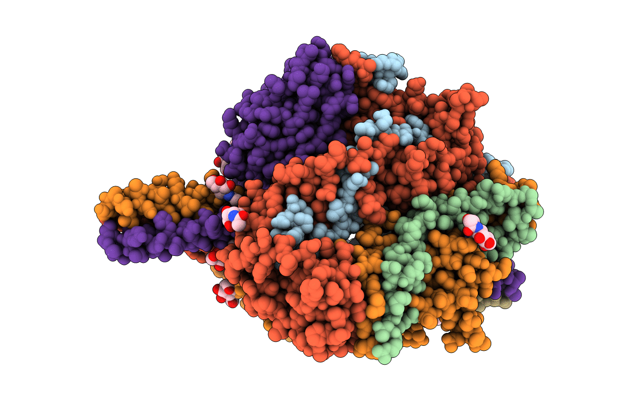
Title:
Structure of the cleavage-activated prefusion form of the parainfluenza virus 5 (PIV5) fusion protein
Biological Source:
Source Organism(s):
Simian virus 5 (Taxon ID: 11208)
Expression System(s):
Method Details:
Experimental Method:
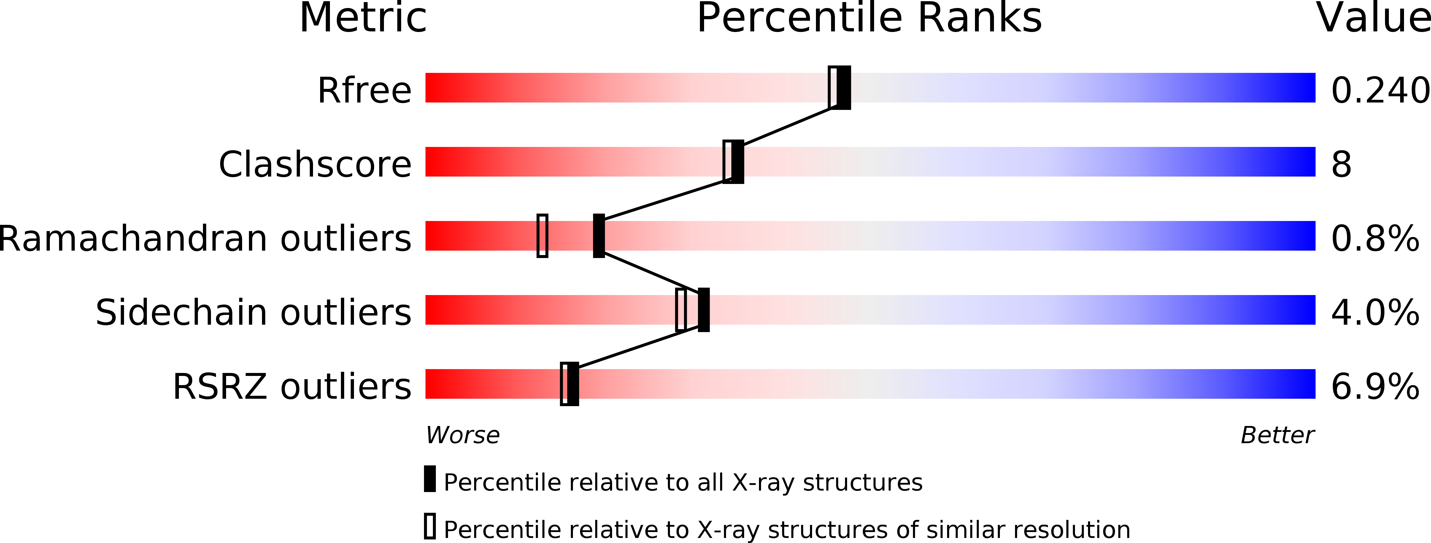
Resolution:
2.00 Å
R-Value Free:
0.24
R-Value Work:
0.22
R-Value Observed:
0.22
Space Group:
C 1 2 1