Deposition Date
2012-08-08
Release Date
2013-06-26
Last Version Date
2024-11-13
Entry Detail
PDB ID:
4GHT
Keywords:
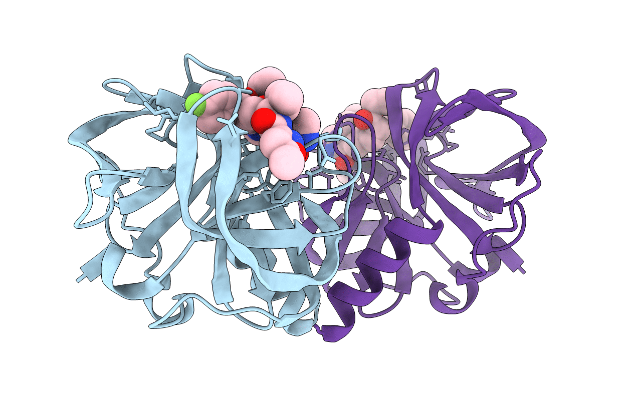
Title:
Crystal structure of EV71 3C proteinase in complex with AG7088
Biological Source:
Source Organism(s):
Human enterovirus 71 (Taxon ID: 39054)
Expression System(s):
Method Details:
Experimental Method:
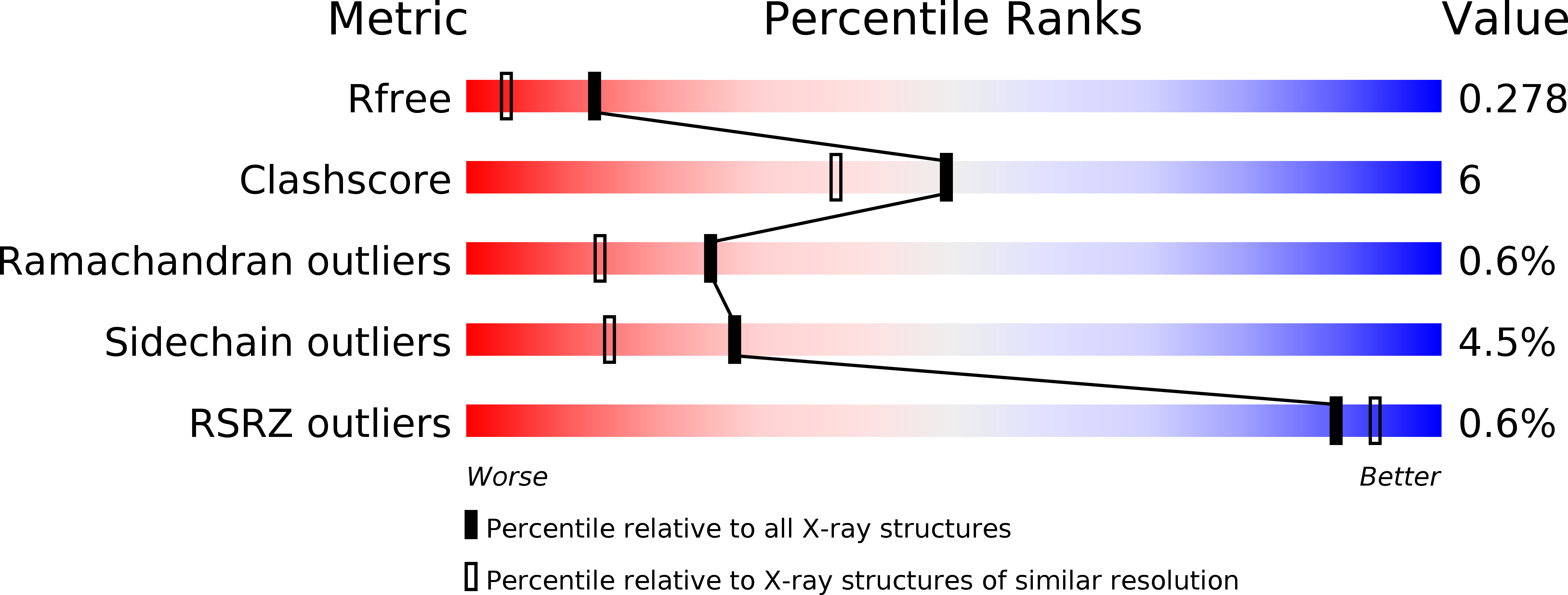
Resolution:
1.96 Å
R-Value Free:
0.27
R-Value Work:
0.20
R-Value Observed:
0.20
Space Group:
P 21 21 21