4GGV image
Deposition Date 2012-08-07
Release Date 2013-07-17
Last Version Date 2023-11-08
Entry Detail
PDB ID:
4GGV
Keywords:
Title:
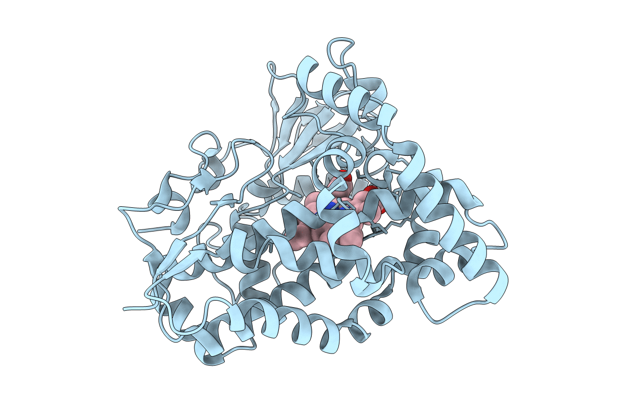
Crystal Structure of HmtT Involved in Himastatin Biosynthesis
Biological Source:
Source Organism:
Host Organism:
Method Details:
Experimental Method:
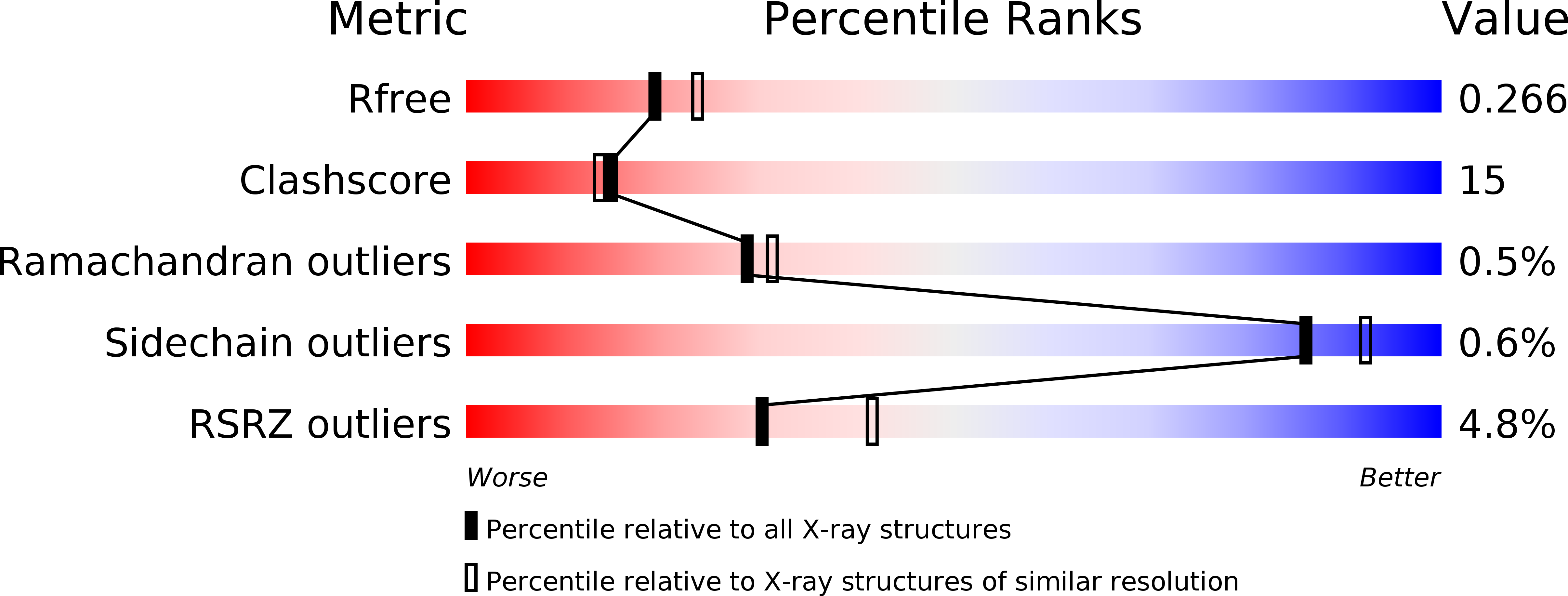
Resolution:
2.33 Å
R-Value Free:
0.25
R-Value Work:
0.20
R-Value Observed:
0.21
Space Group:
P 21 21 2
Macromolecular Entities
Structures with similar UniProt ID
Protein Blast
Polymer Type:polypeptide(L)
Molecule:Cytochrome P450 superfamily protein
Gene (Uniprot):hmtT
Chain IDs:A
Chain Length:418
Number of Molecules:1
Biological Source:Streptomyces himastatinicus
Ligand Molecules
Primary Citation
Structural analysis of HmtT and HmtN involved in the tailoring steps of himastatin biosynthesis
Febs Lett. 587 1675 1680 (2013)
PMID: 23611984 DOI: 10.1016/j.febslet.2013.04.013

Abstact

Himastatin is a novel antibiotic featuring a bicyclohexadepsipeptide structure. On the himastatin biosynthesis pathway, three cytochrome P450s (HmtT, HmtN, HmtS) are responsible for the post-tailoring of the cyclohexadepsipeptide backbone. Here we report the crystal structures of HmtT and HmtN. The overall structures of these two proteins are homologous to other cytochrome P450s. However, the exceptionally long F-G loop in HmtT has a highly unusual conformation and extends deep into the active site. As a result, the F/G helices of HmtT are both kinked. In contrast, the F/G helices of HmtN are straight. Also, the F/G helices in HmtT and HmtN take distinctive orientations, which may be a contributing factor for the substrate specificity of these two enzymes.

Legend

Protein

Chemical

Disease

Primary Citation of related structures