4GD3 image
Entry Detail
PDB ID:
4GD3
Title:
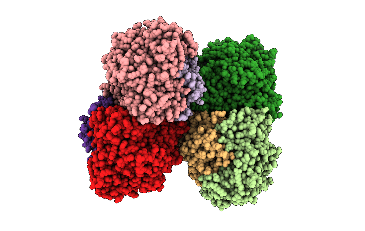
Structure of E. coli hydrogenase-1 in complex with cytochrome b
Biological Source:
Source Organism:
Host Organism:
PDB Version:
Deposition Date:
2012-07-31
Release Date:
2013-01-02
Method Details:
Experimental Method:
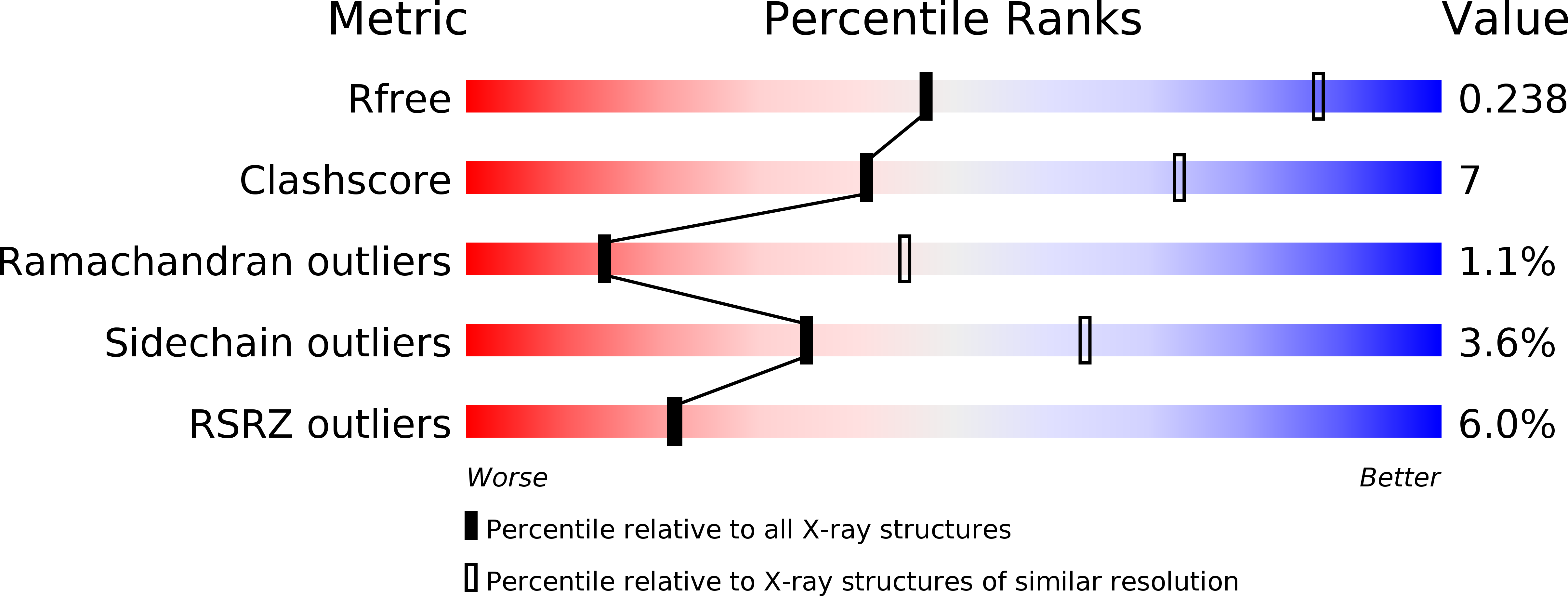
Resolution:
3.30 Å
R-Value Free:
0.23
R-Value Work:
0.19
R-Value Observed:
0.20
Space Group:
P 21 21 21
Macromolecular Entities
Polymer Type:polypeptide(L)
Description:Ni/Fe-hydrogenase 1 B-type cytochrome subunit
Chain IDs:I (auth: A), J (auth: B)
Chain Length:235
Number of Molecules:2
Biological Source:Escherichia coli
Polymer Type:polypeptide(L)
Description:Hydrogenase-1 large chain
Chain IDs:B (auth: L), D (auth: M), F (auth: J), H (auth: K)
Chain Length:582
Number of Molecules:4
Biological Source:Escherichia coli
Polymer Type:polypeptide(L)
Description:Hydrogenase-1 small chain
Mutations:P287C
Chain IDs:A (auth: S), C (auth: T), E (auth: Q), G (auth: R)
Chain Length:335
Number of Molecules:4
Biological Source:Escherichia coli
Primary Citation
Crystal Structure of the O(2)-Tolerant Membrane-Bound Hydrogenase 1 from Escherichia coli in Complex with Its Cognate Cytochrome b.
Structure 21 184 190 (2013)
PMID: 23260654 DOI: 10.1016/j.str.2012.11.010

Abstact

We report the 3.3 Å resolution structure of dimeric membrane-bound O(2)-tolerant hydrogenase 1 from Escherichia coli in a 2:1 complex with its physiological partner, cytochrome b. From the short distance between distal [Fe(4)S(4)] clusters, we predict rapid transfer of H(2)-derived electrons between hydrogenase heterodimers. Thus, under low O(2) levels, a functional active site in one heterodimer can reductively reactivate its O(2)-exposed counterpart in the other. Hydrogenase 1 is maximally expressed during fermentation, when electron acceptors are scarce. These conditions are achieved in the lower part of the host's intestinal tract when E. coli is soon to be excreted and undergo an anaerobic-to-aerobic metabolic transition. The apparent paradox of having an O(2)-tolerant hydrogenase expressed under anoxia makes sense if the enzyme functions to keep intracellular O(2) levels low by reducing it to water, protecting O(2)-sensitive enzymes during the transition. Cytochrome b's main role may be anchoring the hydrogenase to the membrane.

Legend

Protein

Chemical

Disease

Primary Citation of related structures