4GBT image
Deposition Date 2012-07-27
Release Date 2013-02-27
Last Version Date 2024-02-28
Entry Detail
PDB ID:
4GBT
Keywords:
Title:
Structural characterization of H-1 Parvovirus: comparison of infectious virions to replication defective particles
Biological Source:
Source Organism:
H-1 parvovirus (Taxon ID: 10799)
Method Details:
Experimental Method:
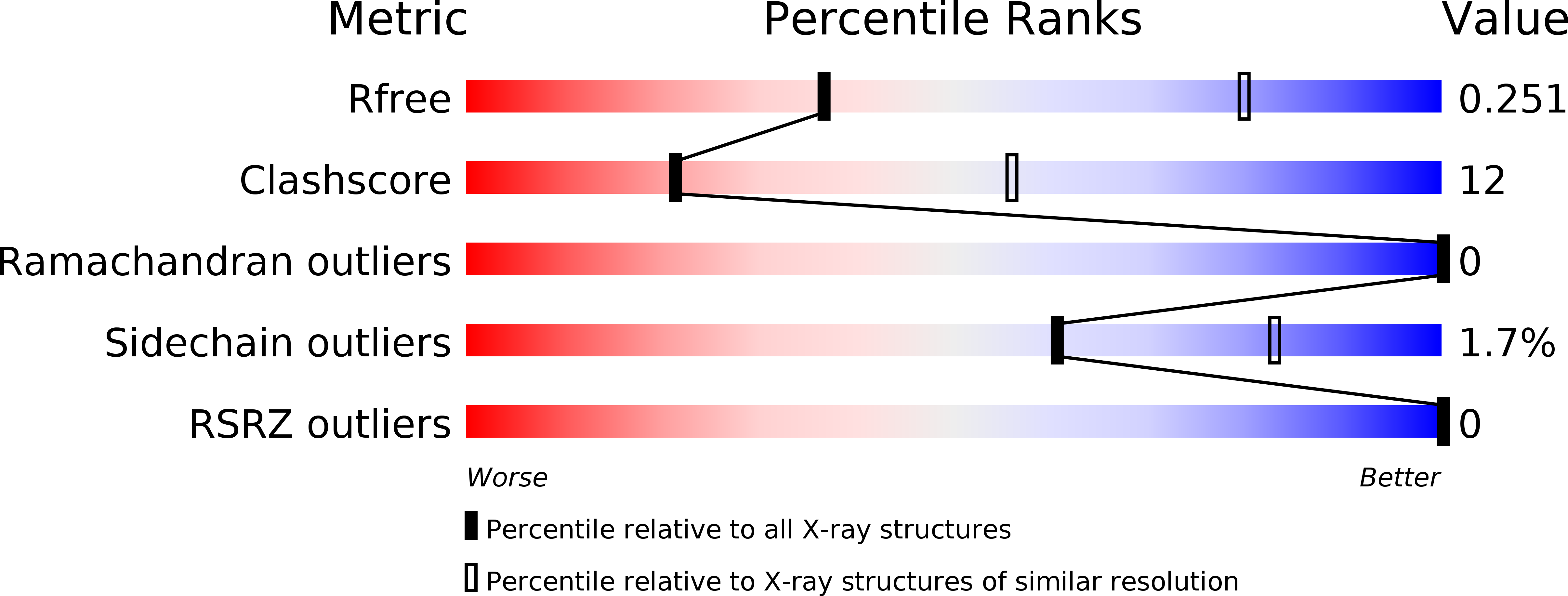
Resolution:
3.20 Å
R-Value Free:
0.25
R-Value Work:
0.25
Space Group:
P 1 21 1
Macromolecular Entities
Structures with similar UniProt ID
Protein Blast
Polymer Type:polypeptide(L)
Molecule:Capsid protein VP1
Chain IDs:A
Chain Length:735
Number of Molecules:1
Biological Source:H-1 parvovirus
Primary Citation
Structural characterization of h-1 parvovirus: comparison of infectious virions to empty capsids.
J.Virol. 87 5128 5140 (2013)
PMID: 23449783 DOI: 10.1128/JVI.03416-12

Abstact

The structure of single-stranded DNA (ssDNA) packaging H-1 parvovirus (H-1PV), which is being developed as an antitumor gene delivery vector, has been determined for wild-type (wt) virions and noninfectious (empty) capsids to 2.7- and 3.2-Å resolution, respectively, using X-ray crystallography. The capsid viral protein (VP) structure consists of an α-helix and an eight-stranded anti-parallel β-barrel with large loop regions between the strands. The β-barrel and loops form the capsid core and surface, respectively. In the wt structure, 600 nucleotides are ordered in an interior DNA binding pocket of the capsid. This accounts for ∼12% of the H-1PV genome. The wt structure is identical to the empty capsid structure, except for side chain conformation variations at the nucleotide binding pocket. Comparison of the H-1PV nucleotides to those observed in canine parvovirus and minute virus of mice, two members of the genus Parvovirus, showed both similarity in structure and analogous interactions. This observation suggests a functional role, such as in capsid stability and/or ssDNA genome recognition for encapsulation. The VP structure differs from those of other parvoviruses in surface loop regions that control receptor binding, tissue tropism, pathogenicity, and antibody recognition, including VP sequences reported to determine tumor cell tropism for oncotropic rodent parvoviruses. These structures of H-1PV provide insight into structural features that dictate capsid stabilization following genome packaging and three-dimensional information applicable for rational design of tumor-targeted recombinant gene delivery vectors.

Legend

Protein

Chemical

Disease

Primary Citation of related structures