4G9M image
Deposition Date 2012-07-24
Release Date 2013-06-05
Last Version Date 2023-09-13
Entry Detail
PDB ID:
4G9M
Title:
Crystal structure of the Rhizoctonia solani agglutinin
Biological Source:
Source Organism:
Method Details:
Experimental Method:
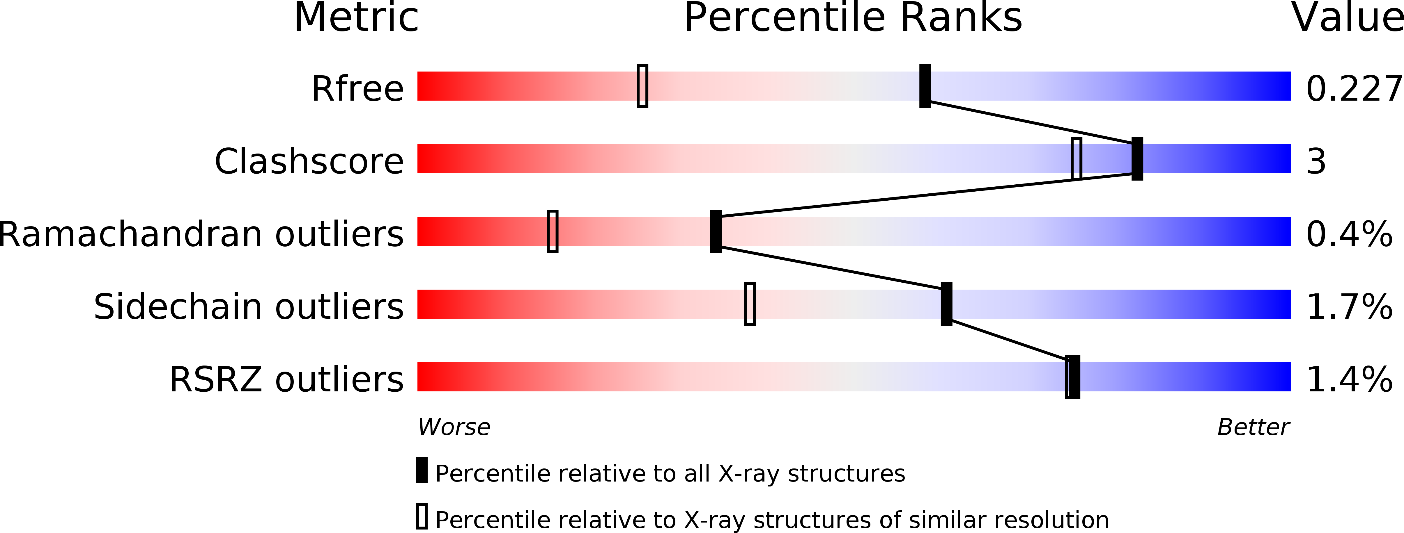
Resolution:
1.60 Å
R-Value Free:
0.23
R-Value Work:
0.18
R-Value Observed:
0.18
Space Group:
C 1 2 1
Macromolecular Entities
Structures with similar UniProt ID
Protein Blast
Polymer Type:polypeptide(L)
Molecule:agglutinin
Chain IDs:A, B
Chain Length:143
Number of Molecules:2
Biological Source:Rhizoctonia solani
Primary Citation
Structural analysis of the Rhizoctonia solani agglutinin reveals a domain-swapping dimeric assembly.
Febs J. 280 1750 1763 (2013)
PMID: 23402398 DOI: 10.1111/febs.12190

Abstact

UNLABELLED Rhizoctonia solani agglutinin (RSA) is a 15.5-kDa lectin accumulated in the mycelium and sclerotia of the soil born plant pathogenic fungus R. solani. Although it is considered to serve as a storage protein and is implicated in fungal insecticidal activity, its physiological role remains unclear as a result of a lack of any structure/function relationship information. Glycan arrays showed that RSA displays high selectivity towards terminal nonreducing N-acetylgalactosamine residues. We determined the amino acid sequence of RSA and also determined the crystal structures of the free form and the RSA-N-acetylgalactosamine complex at 1.6 and 2.2 Å resolution, respectively. RSA is a homodimer comprised of two monomers adopting the β-trefoil fold. Each monomer accommodates two different carbohydrate-binding sites in an asymmetric way. Despite RSA topology similarities with R-type lectins, the two-monomer assembly involves an N-terminal swap, thus creating a dimer association novel to R-type lectins. Structural characterization of the two carbohydrate-binding sites offers insights on the structural determinants of the RSA carbohydrate specificity. DATABASE Structural data have been deposited in the Protein Data Bank database under accession numbers 4G9M and 4G9N. STRUCTURED DIGITAL ABSTRACT RSA and RSA bind by x-ray crystallography (View interaction).

Legend

Protein

Chemical

Disease

Primary Citation of related structures
Feedback Form
Name
Email
Institute
Feedback