4G86 image
Deposition Date 2012-07-21
Release Date 2012-10-24
Last Version Date 2024-02-28
Entry Detail
PDB ID:
4G86
Keywords:
Title:
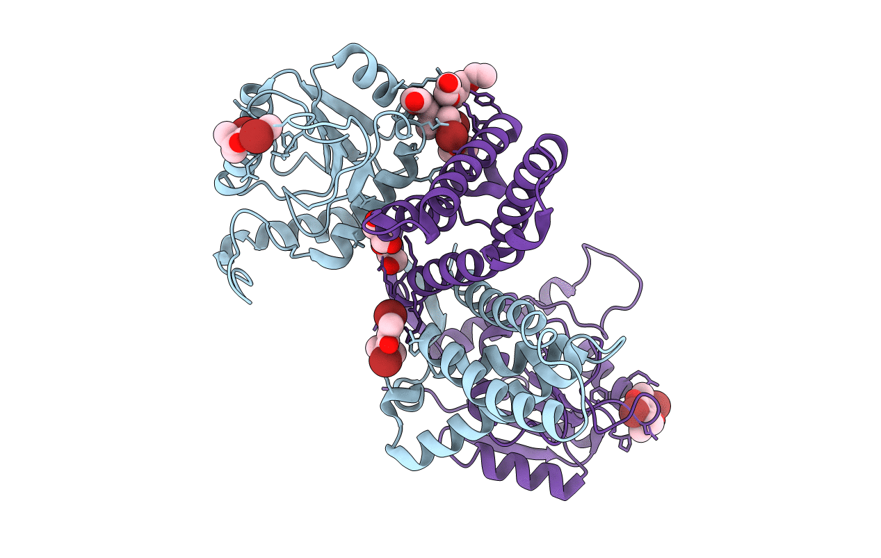
Crystal structure of the redox-active cofactor DBMIB bound to the full length circadian clock protein KaiA from Synechococcus elongatus
Biological Source:
Source Organism:
Host Organism:
Method Details:
Experimental Method:
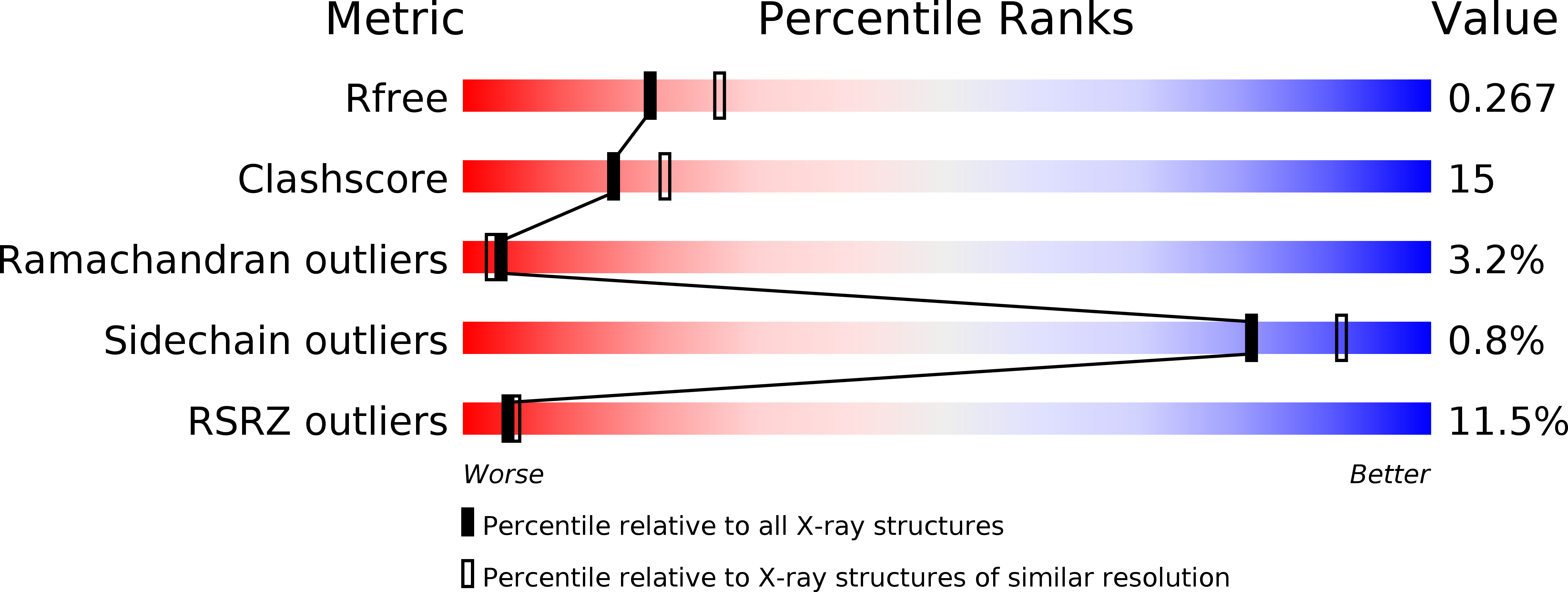
Resolution:
2.39 Å
R-Value Free:
0.26
R-Value Work:
0.21
R-Value Observed:
0.22
Space Group:
P 1 21 1
Macromolecular Entities
Structures with similar UniProt ID
Protein Blast
Polymer Type:polypeptide(L)
Molecule:Circadian clock protein kaiA
Gene (Uniprot):kaiA
Chain IDs:A, B
Chain Length:284
Number of Molecules:2
Biological Source:Synechococcus elongatus
Primary Citation
Crystal Structure of the Redox-Active Cofactor Dibromothymoquinone Bound to Circadian Clock Protein KaiA and Structural Basis for Dibromothymoquinone's Ability to Prevent Stimulation of KaiC Phosphorylation by KaiA.
Biochemistry 51 8050 8052 (2012)
PMID: 23020633 DOI: 10.1021/bi301222t

Abstact

KaiA protein that stimulates KaiC phosphorylation in the cyanobacterial circadian clock was recently shown to be destabilized by dibromothymoquinone (DBMIB), thus revealing KaiA as a sensor of the plastoquinone (PQ) redox state and suggesting an indirect control of the clock by light through PQ redox changes. Here we show using X-ray crystallography that several DBMIBs are bound to KaiA dimer. Some binding modes are consistent with oligomerization of N-terminal KaiA pseudoreceiver domains and/or reduced interdomain flexibility. DBMIB bound to the C-terminal KaiA (C-KaiA) domain and limited stimulation of KaiC kinase activity by C-KaiA in the presence of DBMIB demonstrate that the cofactor may weakly inhibit KaiA-KaiC binding.

Legend

Protein

Chemical

Disease

Primary Citation of related structures
Feedback Form
Name
Email
Institute
Feedback