Deposition Date
2012-07-20
Release Date
2014-02-05
Last Version Date
2024-10-16
Entry Detail
PDB ID:
4G80
Keywords:
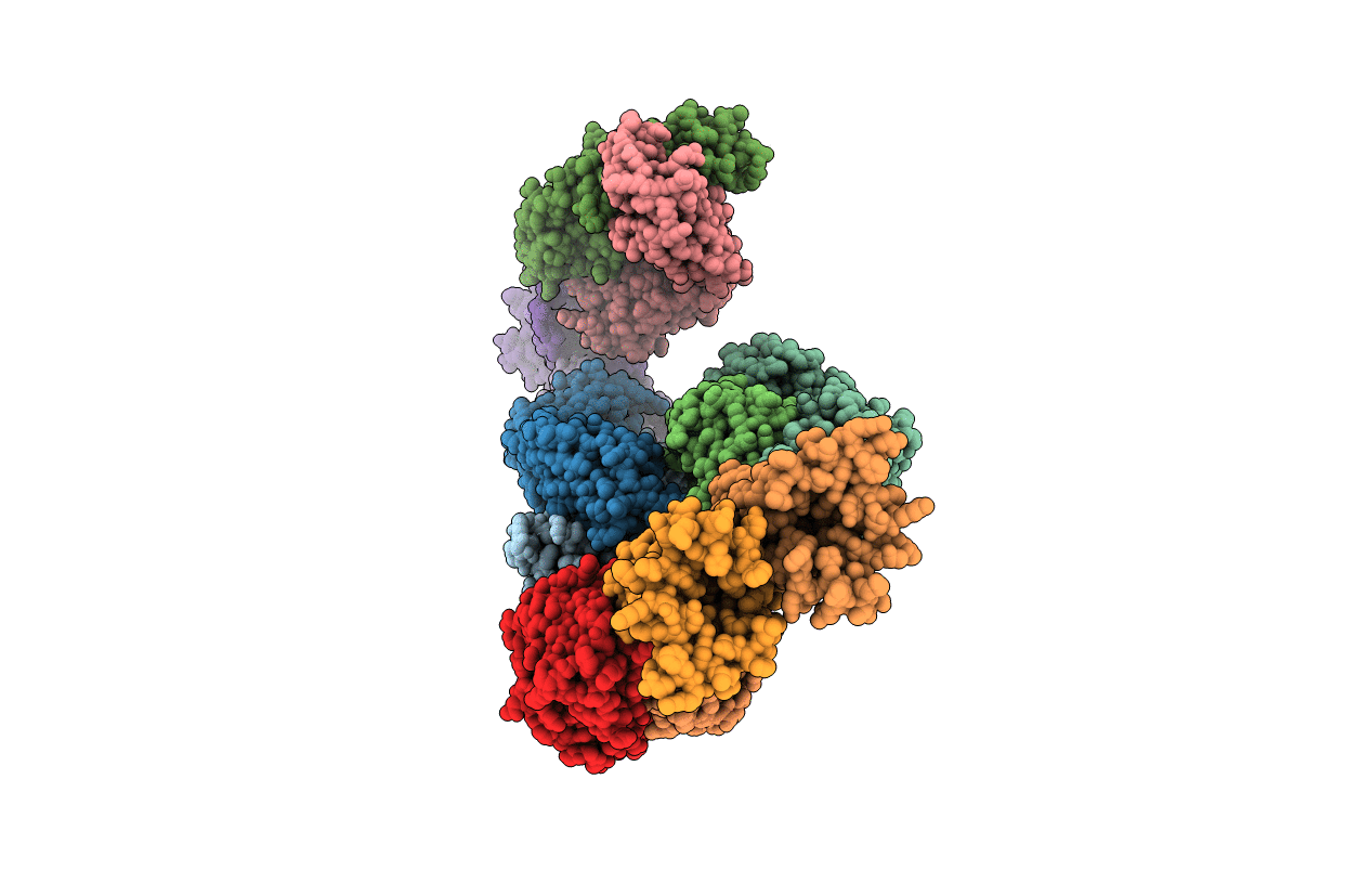
Title:
Crystal structure of voltage sensing domain of Ci-VSP with fragment antibody (WT, 3.8 A)
Biological Source:
Source Organism(s):
Ciona intestinalis (Taxon ID: 7719)
Homo sapiens (Taxon ID: 9606)
Homo sapiens (Taxon ID: 9606)
Expression System(s):
Method Details:
Experimental Method:
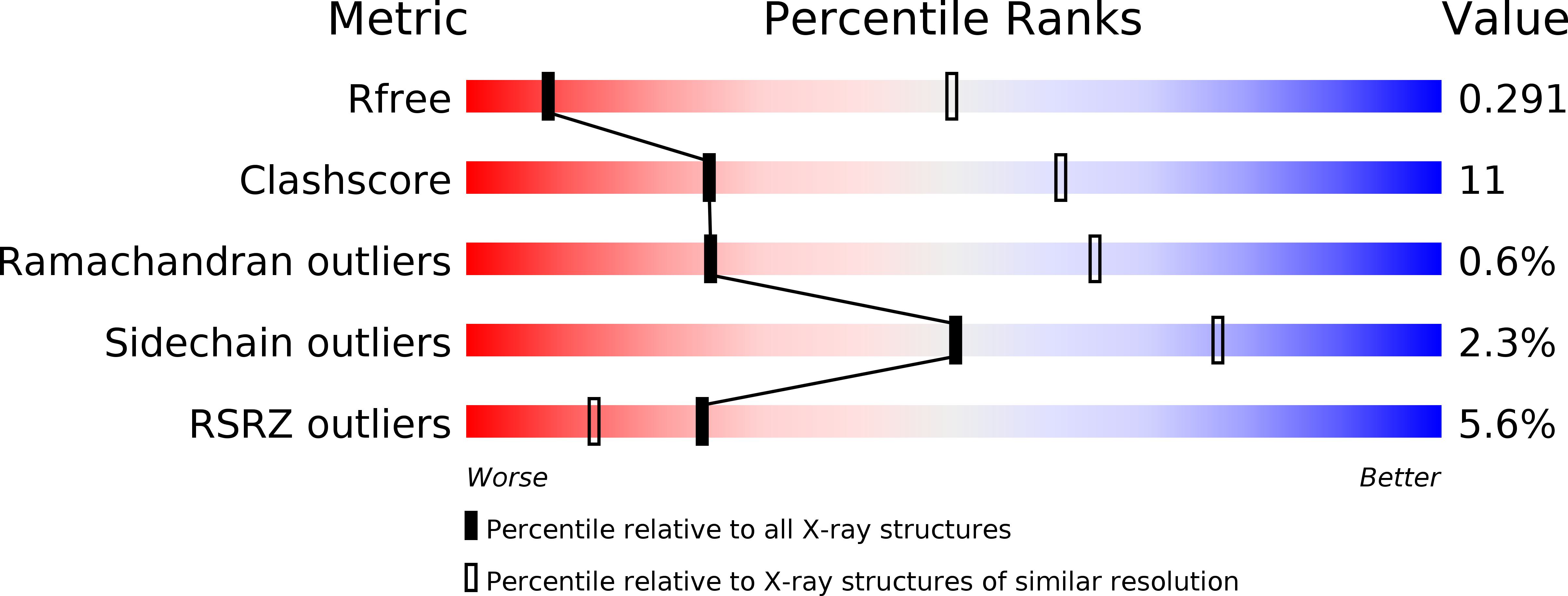
Resolution:
3.58 Å
R-Value Free:
0.29
R-Value Work:
0.24
R-Value Observed:
0.25
Space Group:
P 1