Deposition Date
2012-07-18
Release Date
2012-09-26
Last Version Date
2024-10-30
Entry Detail
PDB ID:
4G6A
Keywords:
Title:
Structure of the Hepatitis C virus envelope glycoprotein E2 antigenic region 412-423 bound to the broadly neutralizing antibody AP33
Biological Source:
Source Organism(s):
Homo sapiens (Taxon ID: 9606)
Hepatitis C virus (Taxon ID: 11103)
Hepatitis C virus (Taxon ID: 11103)
Expression System(s):
Method Details:
Experimental Method:
Resolution:
2.50 Å
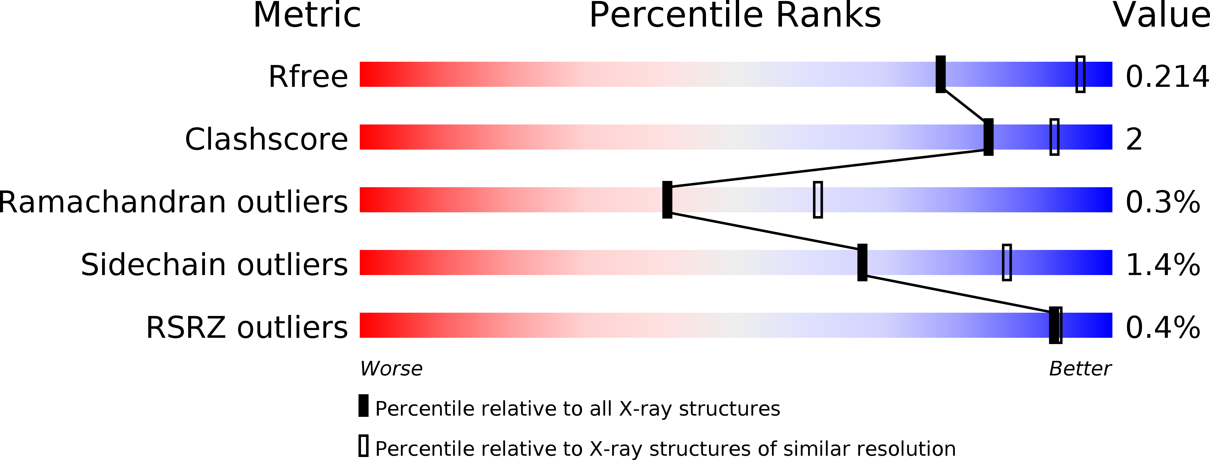
R-Value Free:
0.21
R-Value Work:
0.17
R-Value Observed:
0.18
Space Group:
P 1 21 1