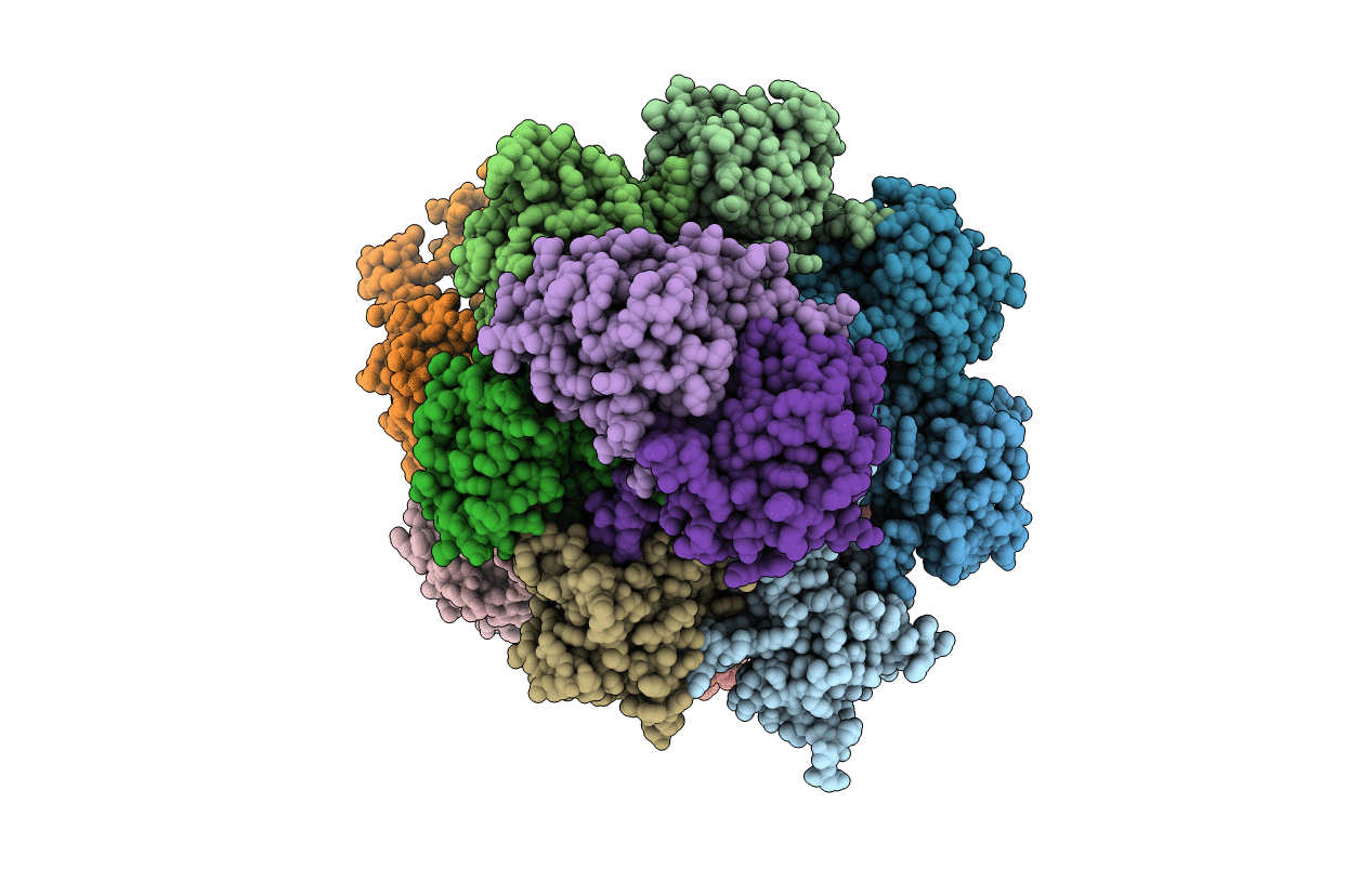
4G4S image
Entry Detail
PDB ID:
4G4S
Title:
Structure of Proteasome-Pba1-Pba2 Complex
Biological Source:
Source Organism:
PDB Version:
Deposition Date:
2012-07-16
Release Date:
2012-09-05
Method Details:
Experimental Method:
Resolution:
2.49 Å
R-Value Free:
0.23
R-Value Work:
0.18
R-Value Observed:
0.18
Space Group:
C 2 2 21
Macromolecular Entities
Polymer Type:polypeptide(L)
Description:Proteasome component C7-alpha
Chain IDs:A
Chain Length:252
Number of Molecules:1
Biological Source:Saccharomyces cerevisiae
Polymer Type:polypeptide(L)
Description:Proteasome component Y7
Chain IDs:B
Chain Length:250
Number of Molecules:1
Biological Source:Saccharomyces cerevisiae
Polymer Type:polypeptide(L)
Description:Proteasome component Y13
Chain IDs:C
Chain Length:258
Number of Molecules:1
Biological Source:Saccharomyces cerevisiae
Polymer Type:polypeptide(L)
Description:Proteasome component PRE6
Chain IDs:D
Chain Length:254
Number of Molecules:1
Biological Source:Saccharomyces cerevisiae
Polymer Type:polypeptide(L)
Description:Proteasome component PUP2
Chain IDs:E
Chain Length:261
Number of Molecules:1
Biological Source:Saccharomyces cerevisiae
Polymer Type:polypeptide(L)
Description:Proteasome component PRE5
Chain IDs:F
Chain Length:235
Number of Molecules:1
Biological Source:Saccharomyces cerevisiae
Polymer Type:polypeptide(L)
Description:Proteasome component C1
Chain IDs:G
Chain Length:288
Number of Molecules:1
Biological Source:Saccharomyces cerevisiae
Polymer Type:polypeptide(L)
Description:Proteasome component PRE3
Chain IDs:H
Chain Length:196
Number of Molecules:1
Biological Source:Saccharomyces cerevisiae
Polymer Type:polypeptide(L)
Description:Proteasome component PUP1
Chain IDs:I
Chain Length:232
Number of Molecules:1
Biological Source:Saccharomyces cerevisiae
Polymer Type:polypeptide(L)
Description:Proteasome component PUP3
Chain IDs:J
Chain Length:205
Number of Molecules:1
Biological Source:Saccharomyces cerevisiae
Polymer Type:polypeptide(L)
Description:Proteasome component C11
Chain IDs:K
Chain Length:198
Number of Molecules:1
Biological Source:Saccharomyces cerevisiae
Polymer Type:polypeptide(L)
Description:Proteasome component PRE2
Chain IDs:L
Chain Length:212
Number of Molecules:1
Biological Source:Saccharomyces cerevisiae
Polymer Type:polypeptide(L)
Description:Proteasome component C5
Chain IDs:M
Chain Length:222
Number of Molecules:1
Biological Source:Saccharomyces cerevisiae
Polymer Type:polypeptide(L)
Description:Proteasome component PRE4
Chain IDs:N
Chain Length:233
Number of Molecules:1
Biological Source:Saccharomyces cerevisiae
Polymer Type:polypeptide(L)
Description:Proteasome chaperone 1
Chain IDs:O
Chain Length:276
Number of Molecules:1
Biological Source:Saccharomyces cerevisiae
Polymer Type:polypeptide(L)
Description:Proteasome assembly chaperone 2
Chain IDs:P
Chain Length:269
Number of Molecules:1
Biological Source:Saccharomyces cerevisiae
Peptide-like Molecules
PRD_000907
Primary Citation
Structure of a Proteasome Pba1-Pba2 Complex: IMPLICATIONS FOR PROTEASOME ASSEMBLY, ACTIVATION, AND BIOLOGICAL FUNCTION.
J.Biol.Chem. 287 37371 37382 (2012)
PMID: 22930756 DOI: 10.1074/jbc.M112.367003

Abstact

The 20S proteasome is an essential, 28-subunit protease that sequesters proteolytic sites within a central chamber, thereby repressing substrate degradation until proteasome activators open the entrance/exit gate. Two established activators, Blm10 and PAN/19S, induce gate opening by binding to the pockets between proteasome α-subunits using C-terminal HbYX (hydrophobic-tyrosine-any residue) motifs. Equivalent HbYX motifs have been identified in Pba1 and Pba2, which function in proteasome assembly. Here, we demonstrate that Pba1-Pba2 proteins form a stable heterodimer that utilizes its HbYX motifs to bind mature 20S proteasomes in vitro and that the Pba1-Pba2 HbYX motifs are important for a physiological function of proteasomes, the maintenance of mitochondrial function. Other factors that contribute to proteasome assembly or function also act in the maintenance of mitochondrial function and display complex genetic interactions with one another, possibly revealing an unexpected pathway of mitochondrial regulation involving the Pba1-Pba2 proteasome interaction. Our determination of a proteasome Pba1-Pba2 crystal structure reveals a Pba1 HbYX interaction that is superimposable with those of known activators, a Pba2 HbYX interaction that is different from those reported previously, and a gate structure that is disrupted but not sufficiently open to allow entry of even small peptides. These findings extend understanding of proteasome interactions with HbYX motifs and suggest multiple roles for Pba1-Pba2 interactions throughout proteasome assembly and function.

Legend

Protein

Chemical

Disease

Primary Citation of related structures