4G4J image
Entry Detail
PDB ID:
4G4J
Keywords:
Title:
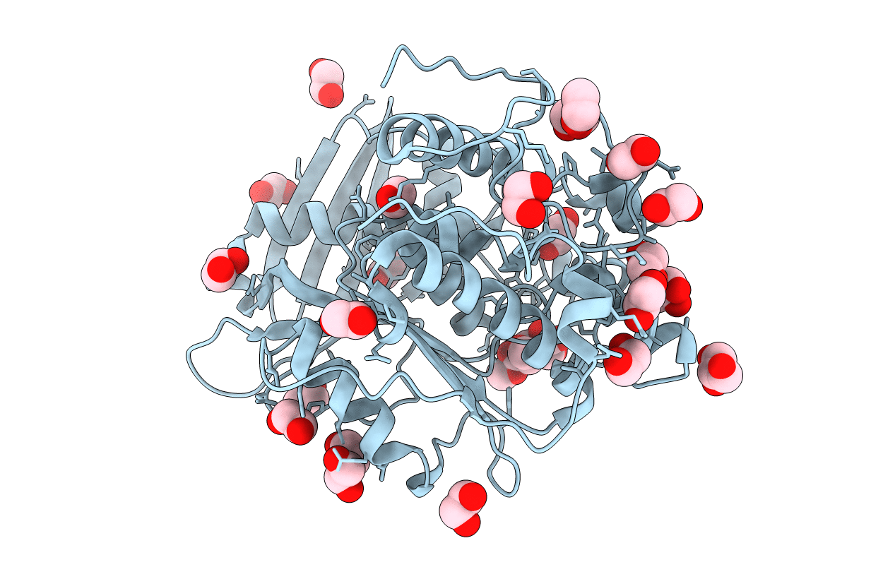
Crystal structure of glucuronoyl esterase S213A mutant from Sporotrichum thermophile in complex with methyl 4-O-methyl-beta-D-glucopyranuronate determined at 2.35 A resolution
Biological Source:
Host Organism:
PDB Version:
Deposition Date:
2012-07-16
Release Date:
2013-01-02
Method Details:
Experimental Method:
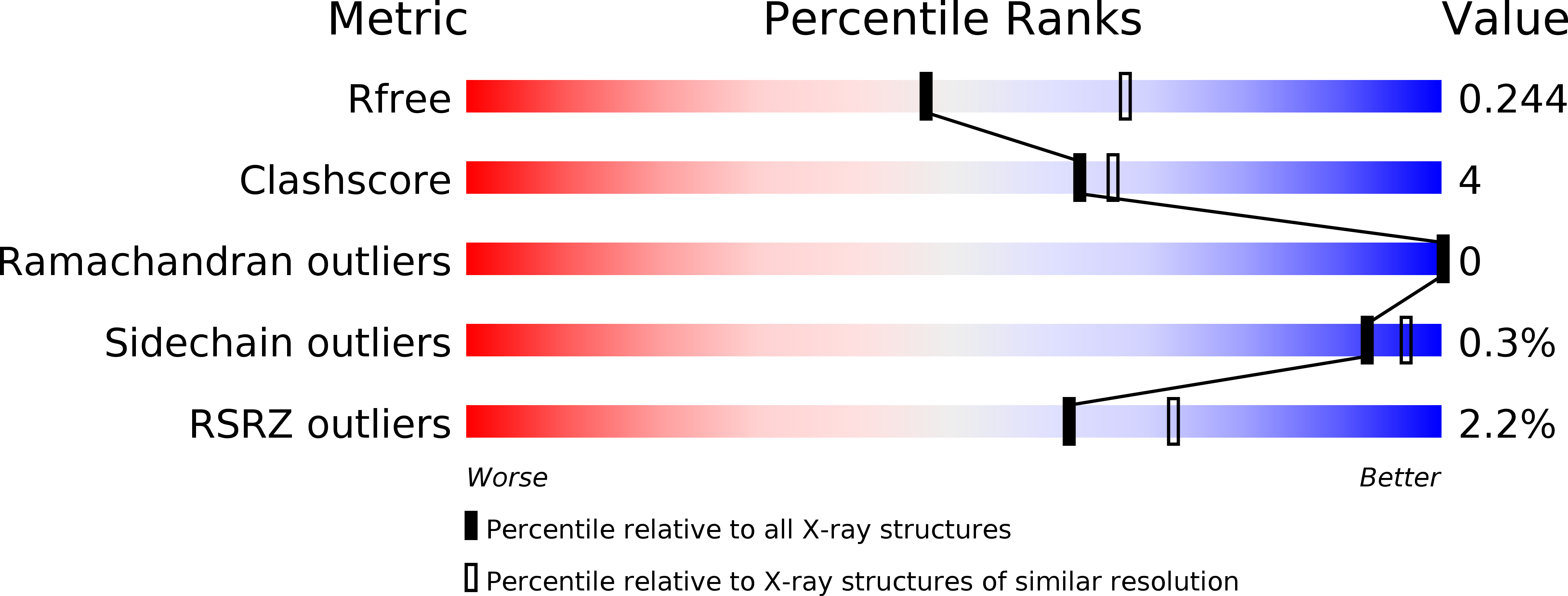
Resolution:
2.35 Å
R-Value Free:
0.24
R-Value Work:
0.18
R-Value Observed:
0.18
Space Group:
P 21 2 21
Macromolecular Entities
Polymer Type:polypeptide(L)
Description:4-O-methyl-glucuronoyl methylesterase
Mutations:S213A
Chain IDs:A
Chain Length:433
Number of Molecules:1
Biological Source:Myceliophthora thermophila
Primary Citation
The structure of a novel glucuronoyl esterase from Myceliophthora thermophila gives new insights into its role as a potential biocatalyst.
Acta Crystallogr.,Sect.D 69 63 73 (2013)
PMID: 23275164 DOI: 10.1107/S0907444912042400

Abstact

The increasing demand for the development of efficient biocatalysts is a consequence of their broad industrial applications. Typical difficulties that are encountered during their exploitation in a variety of processes are interconnected with factors such as temperature, pH, product inhibitors etc. To eliminate these, research has been directed towards the identification of new enzymes that would comply with the required standards. To this end, the recently discovered glucuronoyl esterases (GEs) are an enigmatic family within the carbohydrate esterase (CE) family. Structures of the thermophilic StGE2 esterase from Myceliophthora thermophila (synonym Sporotrichum thermophile), a member of the CE15 family, and its S213A mutant were determined at 1.55 and 1.9 Å resolution, respectively. The first crystal structure of the S213A mutant in complex with a substrate analogue, methyl 4-O-methyl-β-D-glucopyranuronate, was determined at 2.35 Å resolution. All of the three-dimensional protein structures have an α/β-hydrolase fold with a three-layer αβα-sandwich architecture and a Rossmann topology and comprise one molecule per asymmetric unit. These are the first crystal structures of a thermophilic GE both in an unliganded form and bound to a substrate analogue, thus unravelling the organization of the catalytic triad residues and their neighbours lining the active site. The knowledge derived offers novel insights into the key structural elements that drive the hydrolysis of glucuronic acid esters.

Legend

Protein

Chemical

Disease

Primary Citation of related structures