4G3K image
Entry Detail
PDB ID:
4G3K
Title:
Crystal structure of a. aeolicus nlh1 gaf domain in an inactive state
Biological Source:
Source Organism:
Host Organism:
PDB Version:
Deposition Date:
2012-07-14
Release Date:
2013-05-29
Method Details:
Experimental Method:
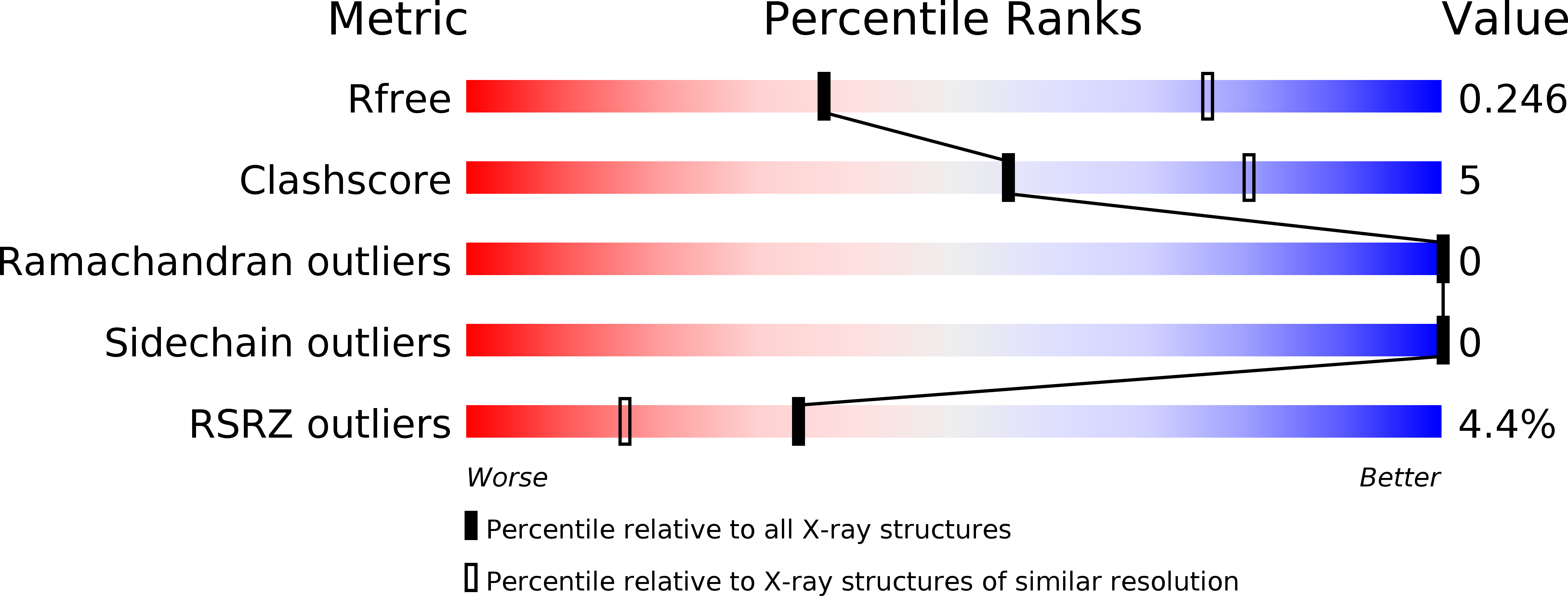
Resolution:
3.05 Å
R-Value Free:
0.24
R-Value Work:
0.20
R-Value Observed:
0.21
Space Group:
P 4 21 2
Macromolecular Entities
Polymer Type:polypeptide(L)
Description:Transcriptional regulator nlh1
Chain IDs:A, B
Chain Length:175
Number of Molecules:2
Biological Source:Aquifex aeolicus
Primary Citation
Structural mechanism of GAF-regulated delta(54) activators from Aquifex aeolicus
J.Mol.Biol. 425 156 170 (2013)
PMID: 23123379 DOI: 10.1016/j.jmb.2012.10.017

Abstact

The σ subunits of bacterial RNA polymerase occur in many variant forms and confer promoter specificity to the holopolymerase. Members of the σ(54) family of σ subunits require the action of a 'transcriptional activator' protein to open the promoter and initiate transcription. The activator proteins undergo regulated assembly from inactive dimers to hexamers that are active ATPases. These contact σ(54) directly and, through ATP hydrolysis, drive a conformational change that enables promoter opening. σ(54) activators use several different kinds of regulatory domains to respond to a wide variety of intracellular signals. One common regulatory module, the GAF domain, is used by σ(54) activators to sense small-molecule ligands. The structural basis for GAF domain regulation in σ(54) activators has not previously been reported. Here, we present crystal structures of GAF regulatory domains for Aquifex aeolicus σ(54) activators NifA-like homolog (Nlh)2 and Nlh1 in three functional states-an 'open', ATPase-inactive state; a 'closed', ATPase-inactive state; and a 'closed', ligand-bound, ATPase-active state. We also present small-angle X-ray scattering data for Nlh2-linked GAF-ATPase domains in the inactive state. These GAF domain dimers regulate σ(54) activator proteins by holding the ATPase domains in an inactive dimer conformation. Ligand binding of Nlh1 dramatically remodels the GAF domain dimer interface, disrupting the contacts with the ATPase domains. This mechanism has strong parallels to the response to phosphorylation in some two-component regulated σ(54) activators. We describe a structural mechanism of GAF-mediated enzyme regulation that appears to be conserved among humans, plants, and bacteria.

Legend

Protein

Chemical

Disease

Primary Citation of related structures