4G38 image
Deposition Date 2012-07-13
Release Date 2012-12-26
Last Version Date 2024-02-28
Entry Detail
PDB ID:
4G38
Keywords:
Title:
Mutational analysis of sulfite reductase hemoprotein reveals the mechanism for coordinated electron and proton transfer
Biological Source:
Source Organism:
Host Organism:
Method Details:
Experimental Method:
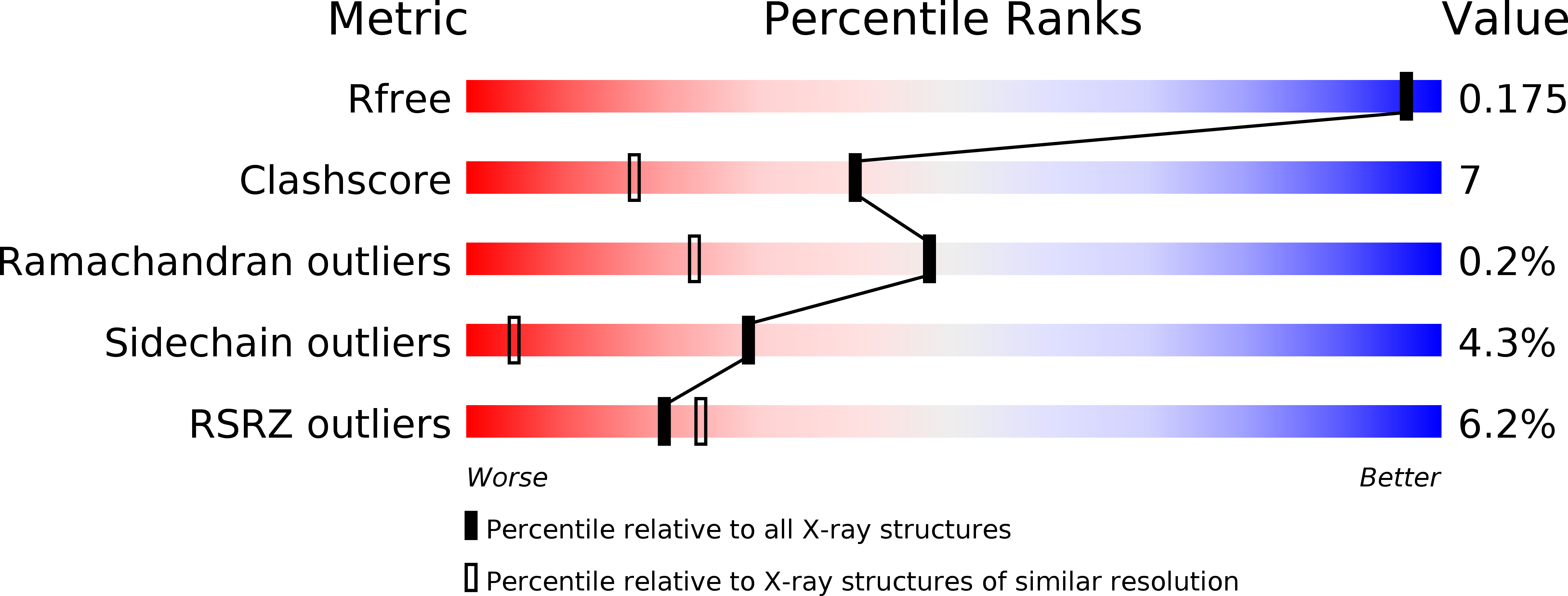
Resolution:
1.56 Å
R-Value Free:
0.17
R-Value Work:
0.14
R-Value Observed:
0.14
Space Group:
P 21 21 21
Macromolecular Entities
Structures with similar UniProt ID
Protein Blast
Polymer Type:polypeptide(L)
Molecule:Sulfite reductase [NADPH] hemoprotein beta-component
Gene (Uniprot):cysI
Mutagens:N149W
Chain IDs:A
Chain Length:570
Number of Molecules:1
Biological Source:Escherichia coli
Primary Citation
Mutational analysis of sulfite reductase hemoprotein reveals the mechanism for coordinated electron and proton transfer.
Biochemistry 51 9857 9868 (2012)
PMID: 23153334 DOI: 10.1021/bi300947a

Abstact

Sulfite reductase catalyzes the six-electron reduction of sulfite to sulfide. The active site, found in the hemoprotein subunit (SiRHP), sits on the distal face of a negatively charged porphyrinoid called siroheme whose central iron atom is coupled to a proximal Fe(4)S(4) cluster. Four positively charged amino acids are positioned around the active site cavity. Together, these two arginines (R83 and R153) and two lysines (K215 and K217) mitigate the negative charge on the siroheme macrocycle. They also serve as a cage around the distally bound anion that tightens when substrate binds and an active site loop clamps down. Structures of native SiRHP point to these amino acids as being important, but their specific roles are ill-defined. Here, we have altered those four active site amino acids and one amino acid on the flexible loop (N149) to probe their roles in SiRHP activity. None of these positively charged residues is required for electron transfer, but only R83S and N149W variants can produce a fully reduced product. By measuring the electrons used per unit of reduced sulfur released, we show that K215, R153, and K217 are responsible for intermediate and late proton transfers, whereas N149 and R153 play a role in the structure of the flexible loop that controls anion binding and release. R83 is primarily responsible for siroheme binding. Together, the activities and structures of these variants reveal specific roles for each in anion binding and in coupled proton transfer that facilitates electron transfer.

Legend

Protein

Chemical

Disease

Primary Citation of related structures
Feedback Form
Name
Email
Institute
Feedback