Deposition Date
2012-07-12
Release Date
2012-09-12
Last Version Date
2024-11-27
Entry Detail
PDB ID:
4G2K
Keywords:
Title:
Crystal structure of the Marburg Virus GP2 ectodomain in its post-fusion conformation
Biological Source:
Source Organism(s):
Saccharomyces cerevisiae (Taxon ID: 4932)
Lake Victoria marburgvirus (Taxon ID: 33728)
Lake Victoria marburgvirus (Taxon ID: 33728)
Expression System(s):
Method Details:
Experimental Method:
Resolution:
1.90 Å
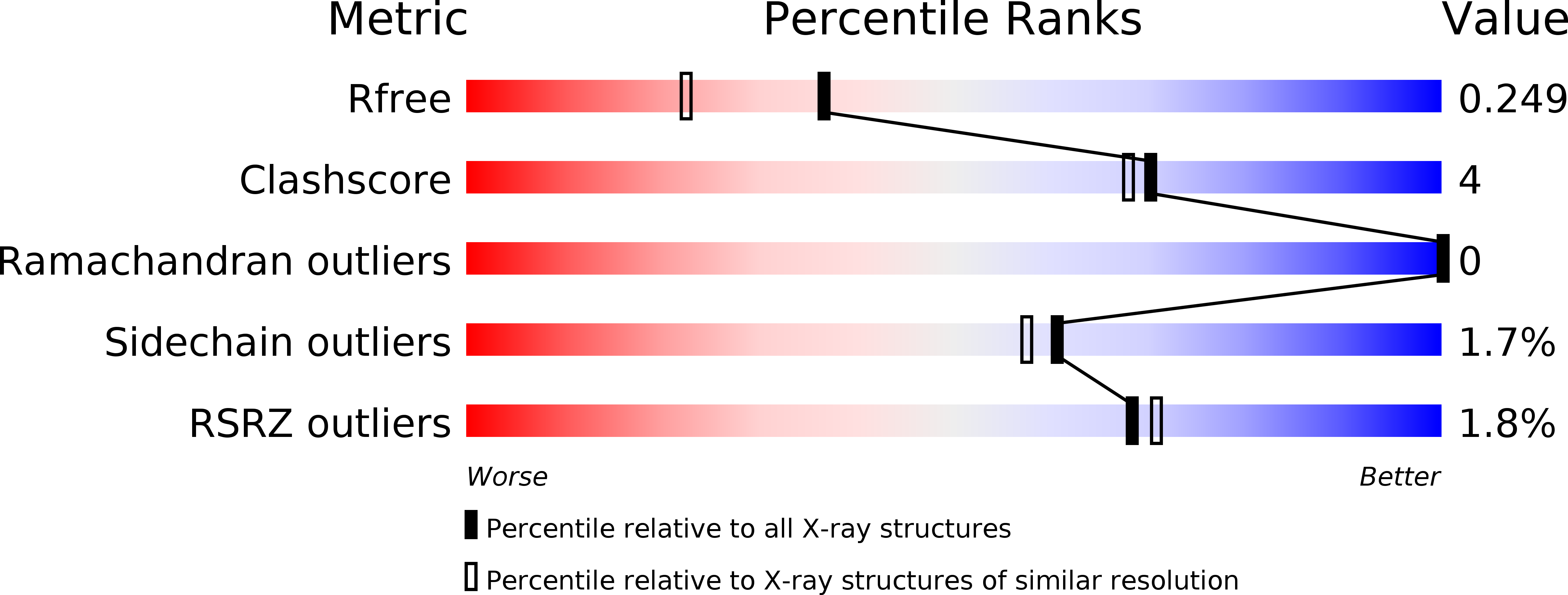
R-Value Free:
0.24
R-Value Work:
0.19
R-Value Observed:
0.19
Space Group:
P 21 21 2