Deposition Date
2012-07-10
Release Date
2012-08-15
Last Version Date
2024-02-28
Entry Detail
PDB ID:
4G1L
Keywords:
Title:
Crystal structure of Newcastle disease virus matrix protein
Biological Source:
Source Organism(s):
Newcastle disease virus (Taxon ID: 11177)
Expression System(s):
Method Details:
Experimental Method:
Resolution:
2.21 Å
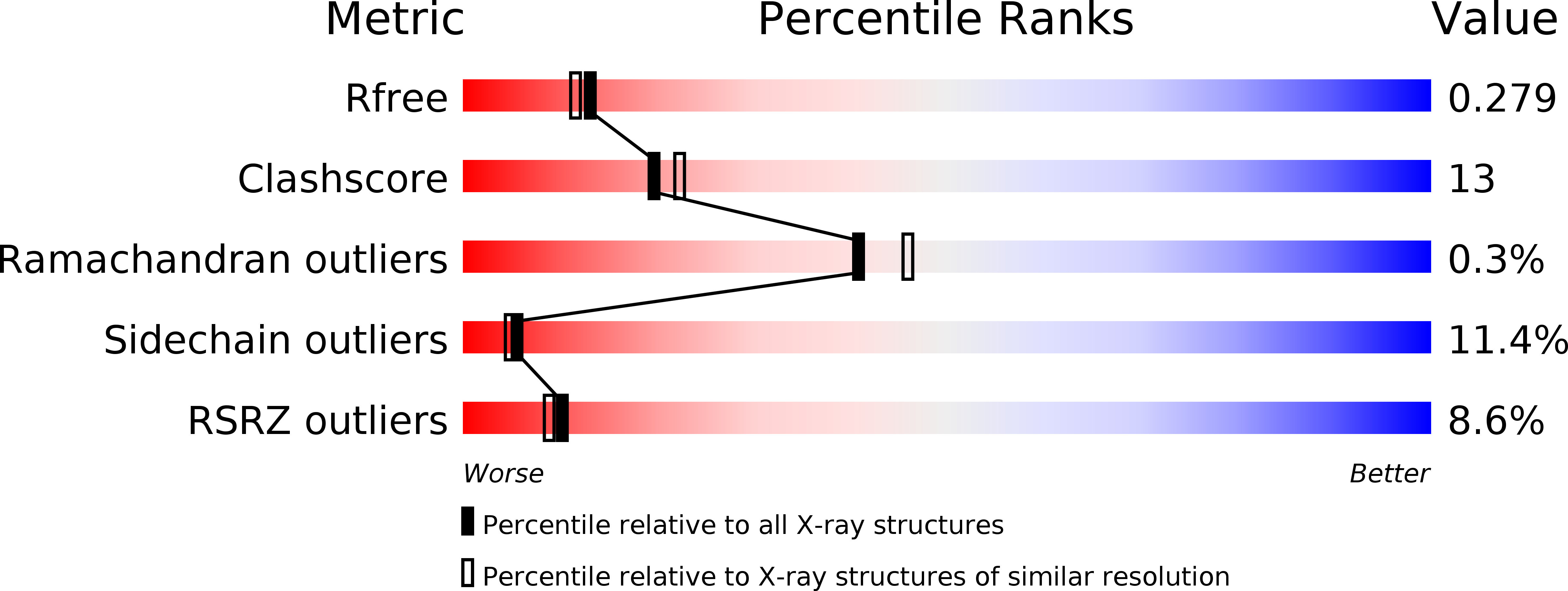
R-Value Free:
0.29
R-Value Work:
0.23
R-Value Observed:
0.23
Space Group:
C 1 2 1