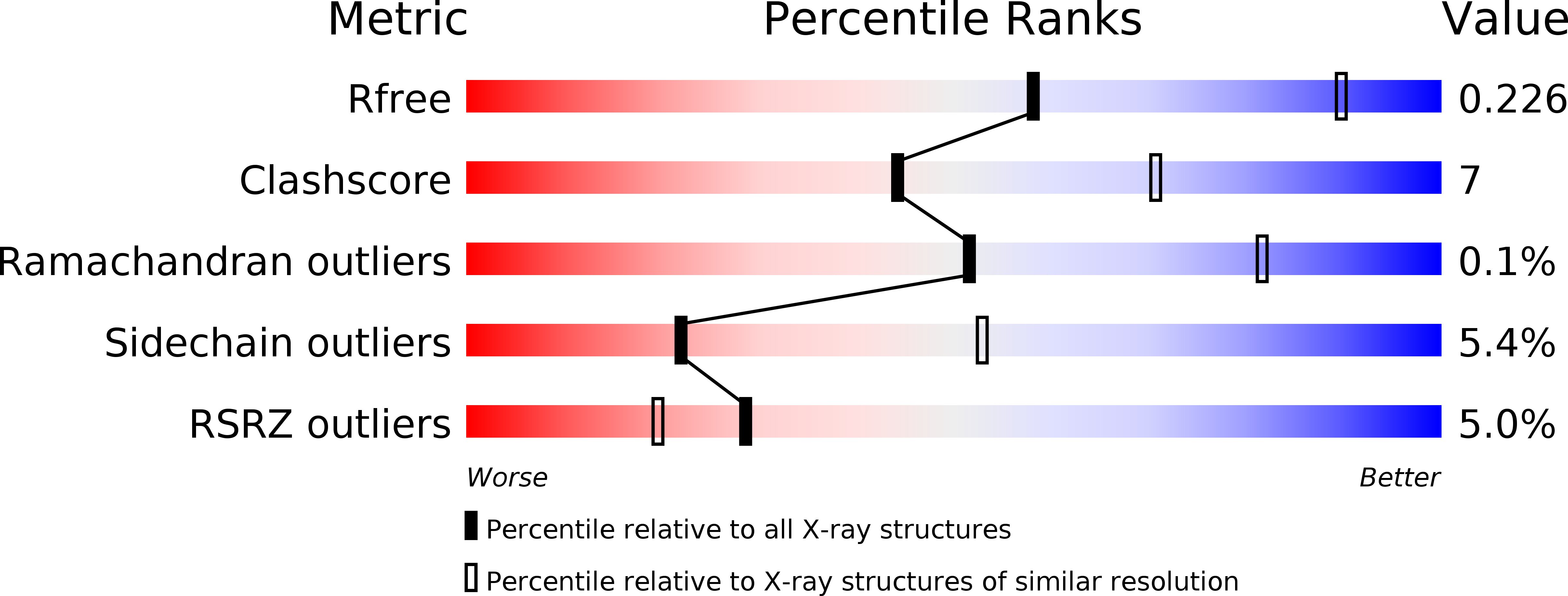
4FZ0 image
Deposition Date 2012-07-05
Release Date 2012-08-01
Last Version Date 2024-11-20
Entry Detail
PDB ID:
4FZ0
Title:
Crystal structure of acid-sensing ion channel in complex with psalmotoxin 1 at low pH
Biological Source:
Source Organism:
Host Organism:
Method Details:
Experimental Method:
Resolution:
2.80 Å
R-Value Free:
0.23
R-Value Work:
0.20
R-Value Observed:
0.20
Space Group:
C 1 2 1
Macromolecular Entities
Polymer Type:polypeptide(L)
Molecule:Acid-sensing ion channel 1
Gene (Uniprot):ASIC1
Chain IDs:A, B, C
Chain Length:450
Number of Molecules:3
Biological Source:Gallus gallus
Polymer Type:polypeptide(L)
Molecule:Pi-theraphotoxin-Pc1a
Chain IDs:D (auth: M), E (auth: N), F (auth: O)
Chain Length:40
Number of Molecules:3
Biological Source:Psalmopoeus cambridgei
Modified Residue
Compound ID Chain ID Parent Comp ID Details 2D Image
ASN B ASN GLYCOSYLATION SITE
Primary Citation
Structural plasticity and dynamic selectivity of acid-sensing ion channel-spider toxin complexes.
Nature 489 400 405 (2012)
PMID: 22842900 DOI: 10.1038/nature11375

Abstact

Acid-sensing ion channels (ASICs) are voltage-independent, amiloride-sensitive channels involved in diverse physiological processes ranging from nociception to taste. Despite the importance of ASICs in physiology, we know little about the mechanism of channel activation. Here we show that psalmotoxin activates non-selective and Na(+)-selective currents in chicken ASIC1a at pH 7.25 and 5.5, respectively. Crystal structures of ASIC1a-psalmotoxin complexes map the toxin binding site to the extracellular domain and show how toxin binding triggers an expansion of the extracellular vestibule and stabilization of the open channel pore. At pH 7.25 the pore is approximately 10 Å in diameter, whereas at pH 5.5 the pore is largely hydrophobic and elliptical in cross-section with dimensions of approximately 5 by 7 Å, consistent with a barrier mechanism for ion selectivity. These studies define mechanisms for activation of ASICs, illuminate the basis for dynamic ion selectivity and provide the blueprints for new therapeutic agents.

Legend

Protein

Chemical

Disease

Primary Citation of related structures