4FWU image
Deposition Date 2012-07-02
Release Date 2012-09-05
Last Version Date 2024-11-06
Entry Detail
PDB ID:
4FWU
Keywords:
Title:
Crystal structure of glutaminyl cyclase from drosophila melanogaster in space group I4
Biological Source:
Source Organism:
Host Organism:
Method Details:
Experimental Method:
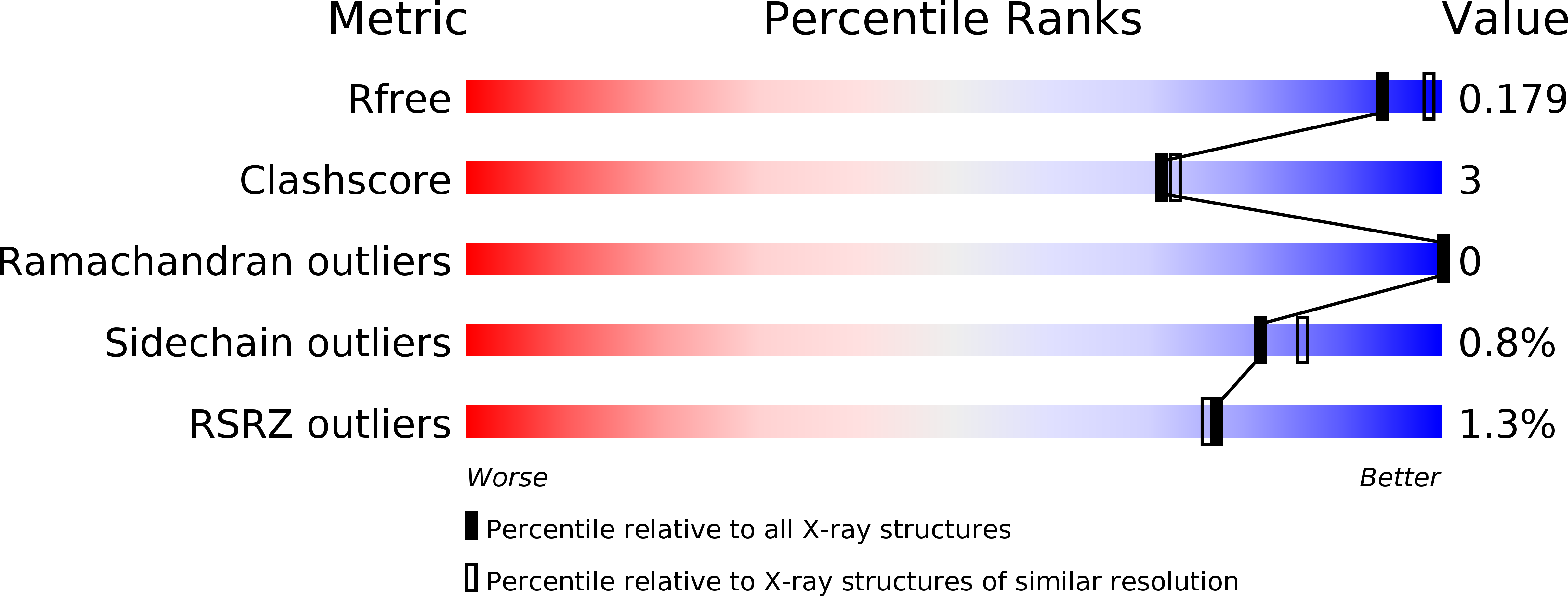
Resolution:
2.00 Å
R-Value Free:
0.20
R-Value Work:
0.16
R-Value Observed:
0.16
Space Group:
I 4
Macromolecular Entities
Structures with similar UniProt ID
Protein Blast
Polymer Type:polypeptide(L)
Molecule:CG32412
Gene (Uniprot):QC
Chain IDs:A
Chain Length:312
Number of Molecules:1
Biological Source:Drosophila melanogaster
Modified Residue
Compound ID Chain ID Parent Comp ID Details 2D Image
ASN A ASN GLYCOSYLATION SITE
Primary Citation
Structure of glutaminyl cyclase from Drosophila melanogaster in space group I4.
Acta Crystallogr.,Sect.F 69 358 361 (2013)
PMID: 23545638 DOI: 10.1107/S1744309113005575

Abstact

The structure of ligand-free glutaminyl cyclase (QC) from Drosophila melanogaster (DmQC) has been determined in a novel crystal form. The protein crystallized in space group I4, with unit-cell parameters a = b = 122.3, c = 72.7 Å. The crystal diffracted to a resolution of 2 Å at the home source. The structure was solved by molecular replacement and was refined to an R factor of 0.169. DmQC exhibits a typical α/β-hydrolase fold. The electron density of three monosaccharides could be localized. The accessibility of the active site will facilitate structural studies of novel inhibitor-binding modes.

Legend

Protein

Chemical

Disease

Primary Citation of related structures