4FVR image
Deposition Date 2012-06-29
Release Date 2012-07-25
Last Version Date 2023-09-13
Entry Detail
PDB ID:
4FVR
Keywords:
Title:
Crystal structure of the Jak2 pseudokinase domain mutant V617F (Mg-ATP-bound form)
Biological Source:
Source Organism:
Homo sapiens (Taxon ID: 9606)
Host Organism:
Method Details:
Experimental Method:
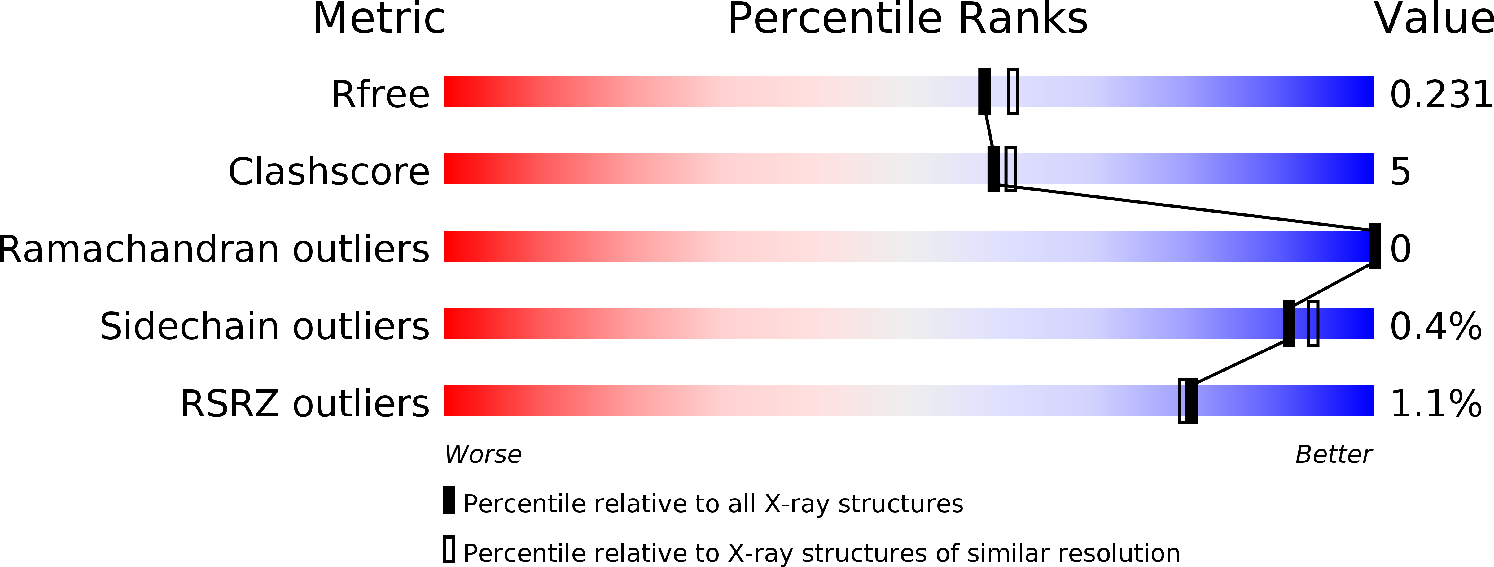
Resolution:
2.00 Å
R-Value Free:
0.22
R-Value Work:
0.18
R-Value Observed:
0.18
Space Group:
P 1 21 1
Macromolecular Entities
Polymer Type:polypeptide(L)
Molecule:Tyrosine-protein kinase JAK2
Gene (Uniprot):JAK2
Mutations:V617F, W777A, F794H
Chain IDs:A
Chain Length:289
Number of Molecules:1
Biological Source:Homo sapiens
Primary Citation
Crystal structures of the JAK2 pseudokinase domain and the pathogenic mutant V617F.
Nat.Struct.Mol.Biol. 19 754 759 (2012)
PMID: 22820988 DOI: 10.1038/nsmb.2348

Abstact

The protein tyrosine kinase JAK2 mediates signaling through numerous cytokine receptors. JAK2 possesses a pseudokinase domain (JH2) and a tyrosine kinase domain (JH1). Through unknown mechanisms, JH2 regulates the catalytic activity of JH1, and hyperactivating mutations in the JH2 region of human JAK2 cause myeloproliferative neoplasms (MPNs). We showed previously that JAK2 JH2 is, in fact, catalytically active. Here we present crystal structures of human JAK2 JH2, including both wild type and the most prevalent MPN mutant, V617F. The structures reveal that JH2 adopts the fold of a prototypical protein kinase but binds Mg-ATP noncanonically. The structural and biochemical data indicate that the V617F mutation rigidifies α-helix C in the N lobe of JH2, facilitating trans-phosphorylation of JH1. The crystal structures of JH2 afford new opportunities for the design of novel JAK2 therapeutics targeting MPNs.

Legend

Protein

Chemical

Disease

Primary Citation of related structures