Deposition Date
2012-06-29
Release Date
2013-06-19
Last Version Date
2024-11-13
Entry Detail
PDB ID:
4FVD
Keywords:
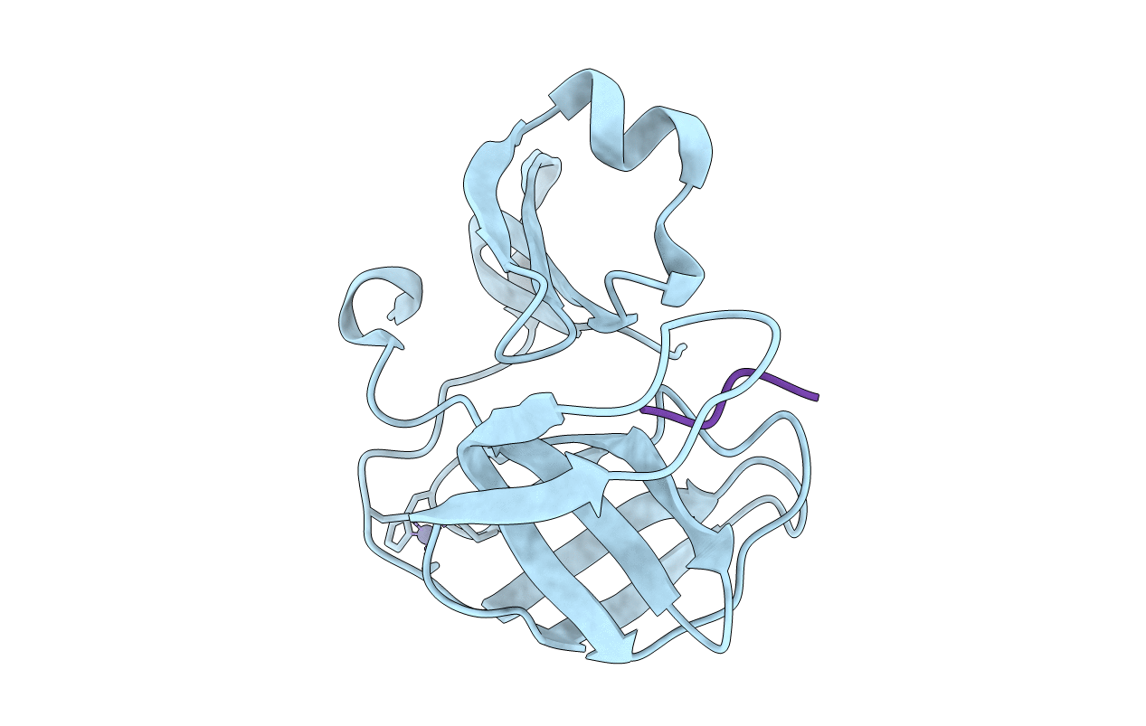
Title:
Crystal structure of EV71 2A proteinase C110A mutant in complex with substrate
Biological Source:
Source Organism(s):
Human enterovirus 71 (Taxon ID: 39054)
Expression System(s):
Method Details:
Experimental Method:
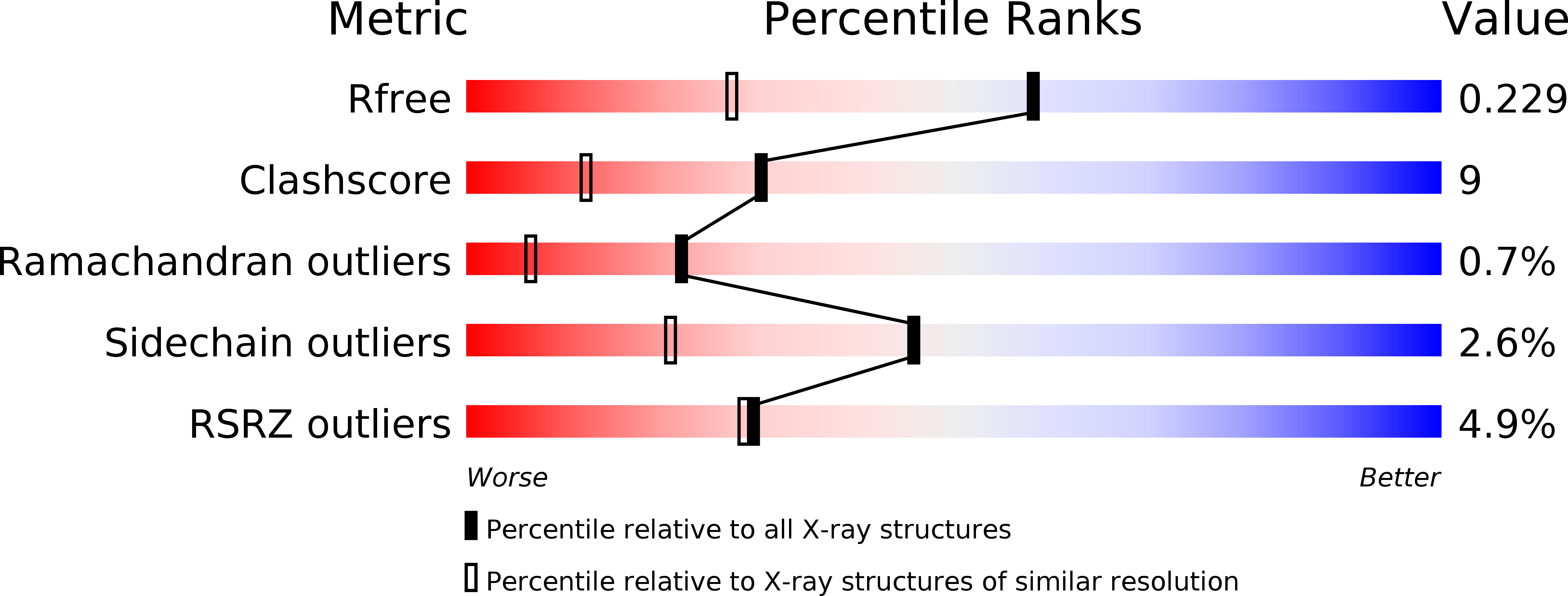
Resolution:
1.66 Å
R-Value Free:
0.22
R-Value Work:
0.18
R-Value Observed:
0.19
Space Group:
C 1 2 1