Deposition Date
2012-06-27
Release Date
2013-06-26
Last Version Date
2023-12-06
Entry Detail
PDB ID:
4FSB
Keywords:
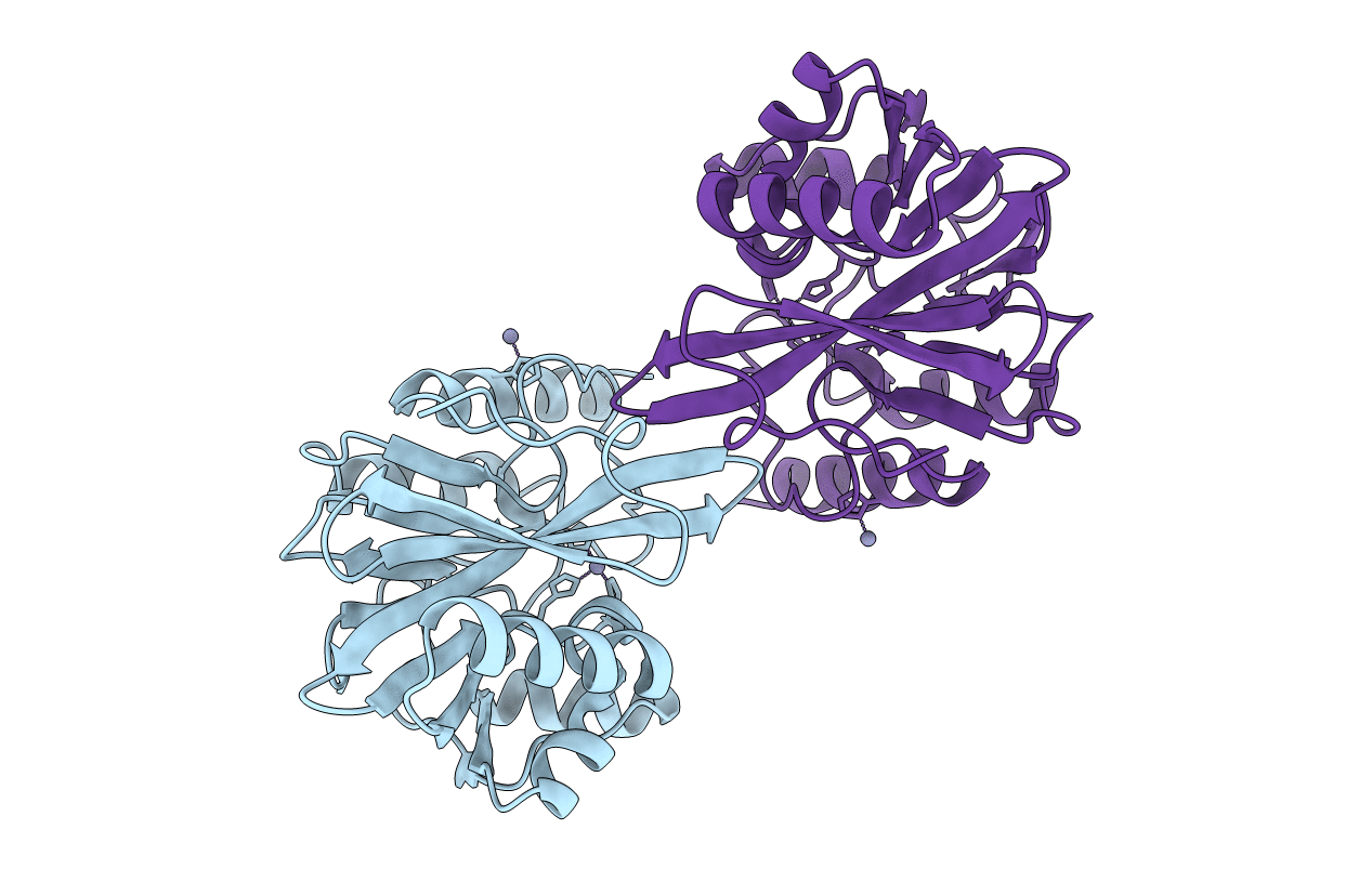
Title:
Crystal structure of the metallo-beta-lactamase VIM-31 in its oxidized form at 1.88 A
Biological Source:
Source Organism(s):
Enterobacter cloacae (Taxon ID: 550)
Expression System(s):
Method Details:
Experimental Method:
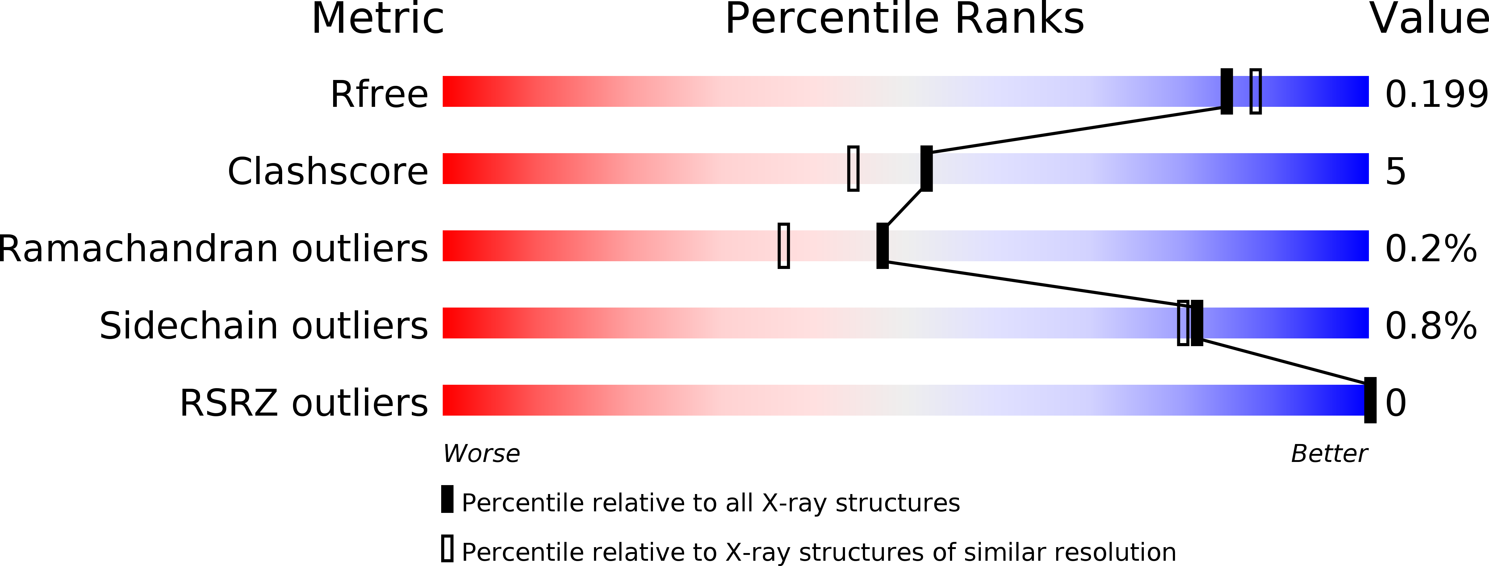
Resolution:
1.88 Å
R-Value Free:
0.19
R-Value Work:
0.14
R-Value Observed:
0.14
Space Group:
I 1 2 1