Deposition Date
2012-06-18
Release Date
2013-02-20
Last Version Date
2023-09-13
Entry Detail
PDB ID:
4FMO
Keywords:
Title:
Structure of the C-terminal domain of the Saccharomyces cerevisiae MUTL alpha (MLH1/PMS1) heterodimer bound to a fragment of exo1
Biological Source:
Source Organism(s):
Saccharomyces cerevisiae (Taxon ID: 559292)
Expression System(s):
Method Details:
Experimental Method:
Resolution:
3.04 Å
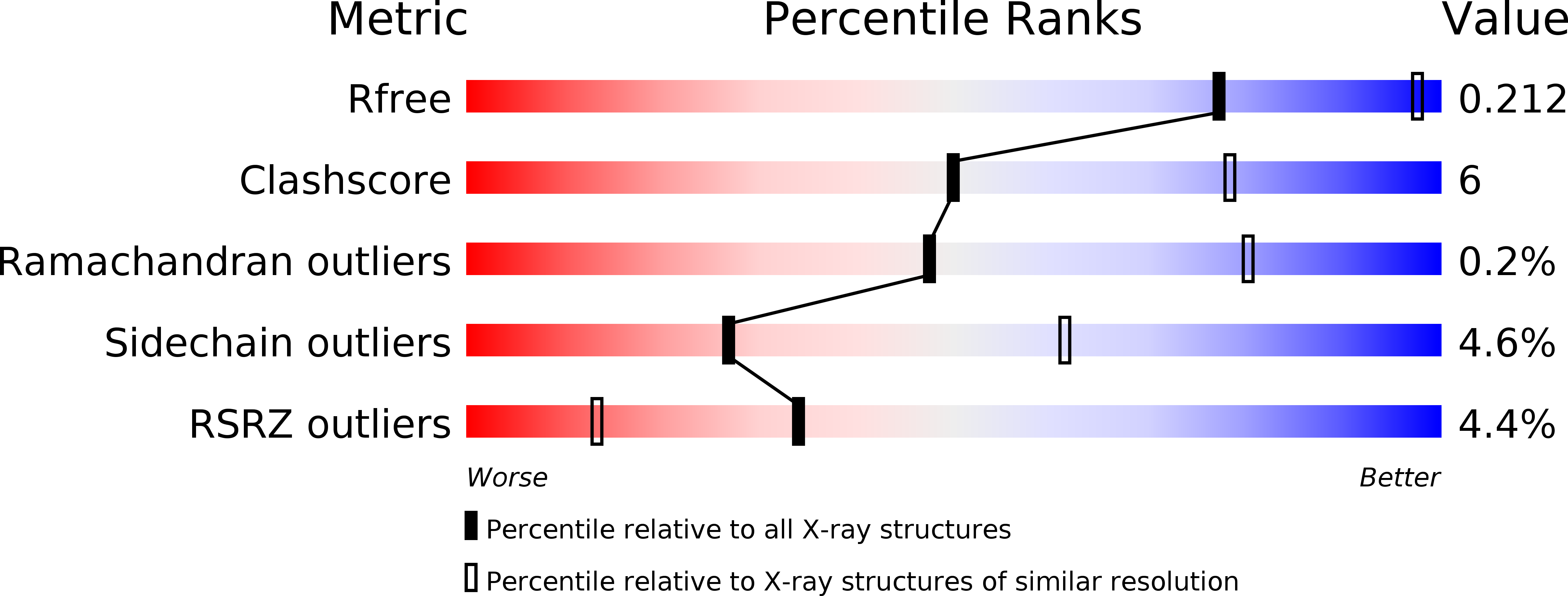
R-Value Free:
0.19
R-Value Work:
0.18
R-Value Observed:
0.18
Space Group:
C 1 2 1