4FKD image
Deposition Date 2012-06-13
Release Date 2013-01-23
Last Version Date 2024-02-28
Entry Detail
PDB ID:
4FKD
Keywords:
Title:
Identification of the Activator Binding Residues in the Second Cysteine-Rich Regulatory Domain of Protein Kinase C Theta
Biological Source:
Source Organism:
Mus musculus (Taxon ID: 10090)
Host Organism:
Method Details:
Experimental Method:
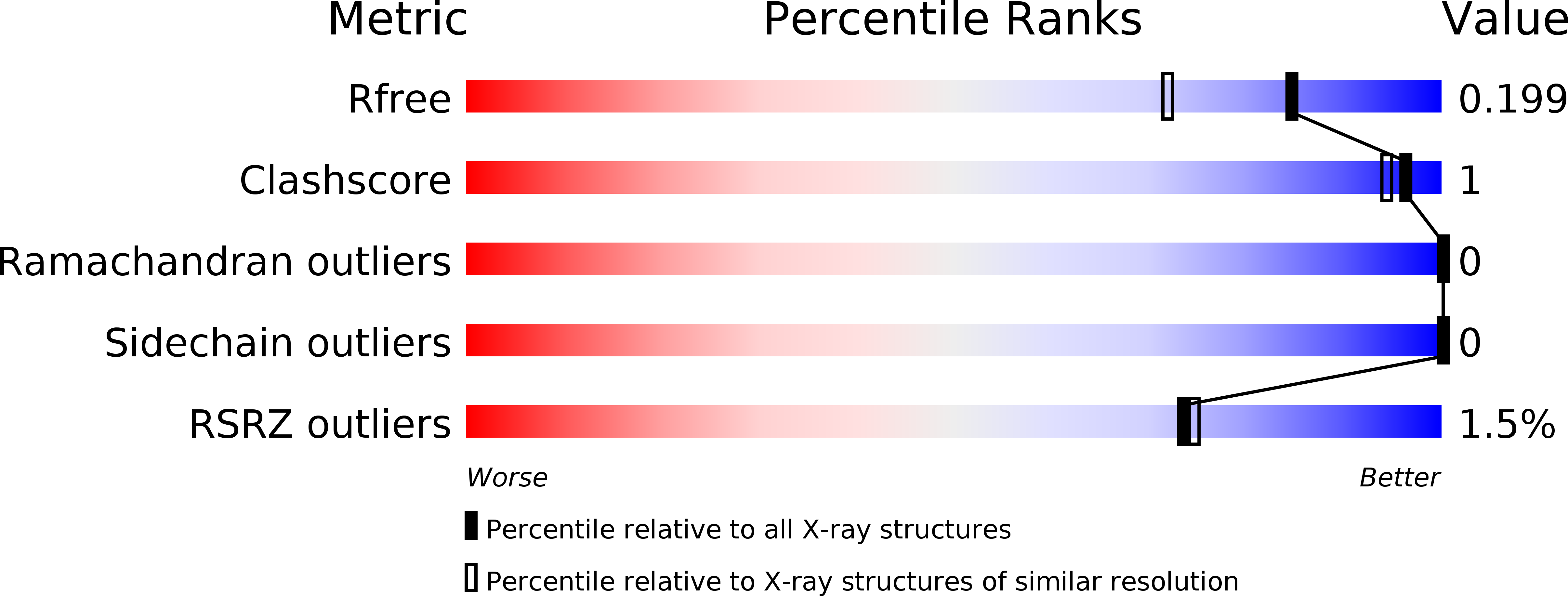
Resolution:
1.63 Å
R-Value Free:
0.20
R-Value Work:
0.17
R-Value Observed:
0.17
Space Group:
P 61
Macromolecular Entities
Structures with similar UniProt ID
Protein Blast
Polymer Type:polypeptide(L)
Molecule:Protein kinase C theta type
Gene (Uniprot):Prkcq
Chain IDs:A
Chain Length:65
Number of Molecules:1
Biological Source:Mus musculus
Ligand Molecules
Primary Citation
Identification of the Activator Binding Residues in the Second Cysteine-Rich Regulatory Domain of Protein Kinase C Theta.
Biochem.J. 451 33 44 (2013)
PMID: 23289588 DOI: 10.1042/BJ20121307

Abstact

PKC (protein kinase C) θ is predominantly expressed in T-cells and is critically involved in immunity. Design of PKCθ-selective molecules to manage autoimmune disorders by targeting its activator-binding C1 domain requires the knowledge of its structure and the activator-binding residues. The C1 domain consists of twin C1 domains, C1A and C1B, of which C1B plays a critical role in the membrane translocation and activation of PKCθ. In the present study we determined the crystal structure of PKCθC1B to 1.63 Å (1 Å=0.1 nm) resolution, which showed that Trp(253) at the rim of the activator-binding pocket was orientated towards the membrane, whereas in PKCδC1B the homologous tryptophan residue was orientated away from the membrane. This particular orientation of Trp(253) affects the size of the activator-binding pocket and the membrane affinity. To further probe the structural constraints on activator-binding, five residues lining the activator-binding site were mutated (Y239A, T243A, W253G, L255G and Q258G) and the binding affinities of the PKCθC1B mutants were measured. These mutants showed reduced binding affinities for phorbol ester [PDBu (phorbol 12,13-dibutyrate)] and diacylglycerol [DOG (sn-1,2-dioctanoylglycerol), SAG (sn-1-stearoyl 2-arachidonyl glycerol)]. All five full-length PKCθ mutants exhibited reduced phorbol-ester-induced membrane translocation compared with the wild-type. These results provide insights into the PKCθ activator-binding domain, which will aid in future design of PKCθ-selective molecules.

Legend

Protein

Chemical

Disease

Primary Citation of related structures