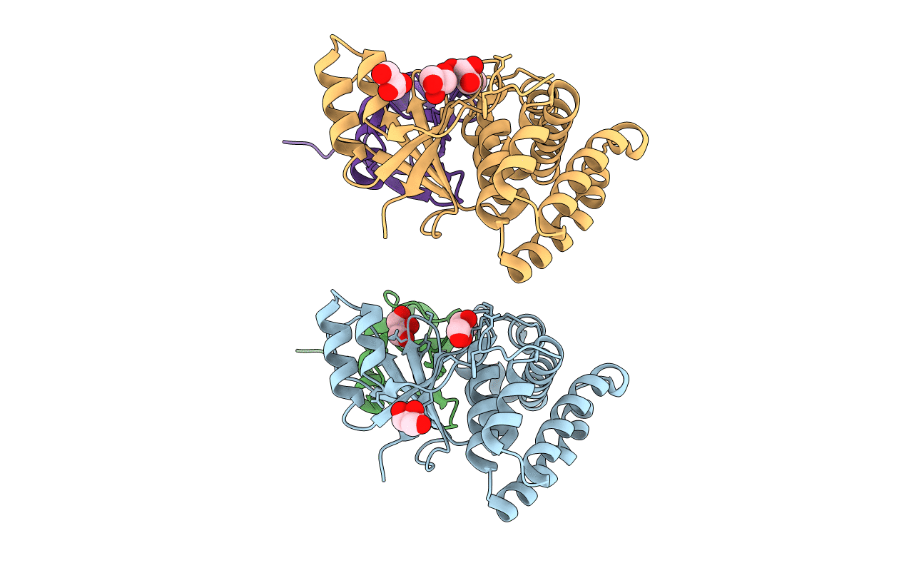
4FJV image
Entry Detail
PDB ID:
4FJV
Keywords:
Title:
Crystal Structure of Human Otubain2 and Ubiquitin Complex
Biological Source:
Source Organism:
Host Organism:
PDB Version:
Deposition Date:
2012-06-12
Release Date:
2013-06-12
Method Details:
Experimental Method:
Resolution:
2.05 Å
R-Value Free:
0.26
R-Value Work:
0.20
R-Value Observed:
0.20
Space Group:
P 21 21 21
Macromolecular Entities
Polymer Type:polypeptide(L)
Description:Ubiquitin thioesterase OTUB2
Chain IDs:A, C
Chain Length:237
Number of Molecules:2
Biological Source:Homo sapiens
Polymer Type:polypeptide(L)
Description:Polyubiquitin-C
Chain IDs:B, D
Chain Length:86
Number of Molecules:2
Biological Source:Homo sapiens
Primary Citation
The human otubain2-ubiquitin structure provides insights into the cleavage specificity of poly-ubiquitin-linkages.
Plos One 10 e0115344 e0115344 (2015)
PMID: 25590432 DOI: 10.1371/journal.pone.0115344

Abstact

Ovarian tumor domain containing proteases cleave ubiquitin (Ub) and ubiquitin-like polypeptides from proteins. Here we report the crystal structure of human otubain 2 (OTUB2) in complex with a ubiquitin-based covalent inhibitor, Ub-Br2. The ubiquitin binding mode is oriented differently to how viral otubains (vOTUs) bind ubiquitin/ISG15, and more similar to yeast and mammalian OTUs. In contrast to OTUB1 which has exclusive specificity towards Lys48 poly-ubiquitin chains, OTUB2 cleaves different poly-Ub linked chains. N-terminal tail swapping experiments between OTUB1 and OTUB2 revealed how the N-terminal structural motifs in OTUB1 contribute to modulating enzyme activity and Ub-chain selectivity, a trait not observed in OTUB2, supporting the notion that OTUB2 may affect a different spectrum of substrates in Ub-dependent pathways.

Legend

Protein

Chemical

Disease

Primary Citation of related structures