4FIW image
Deposition Date 2012-06-11
Release Date 2013-02-06
Last Version Date 2024-11-06
Entry Detail
PDB ID:
4FIW
Keywords:
Title:
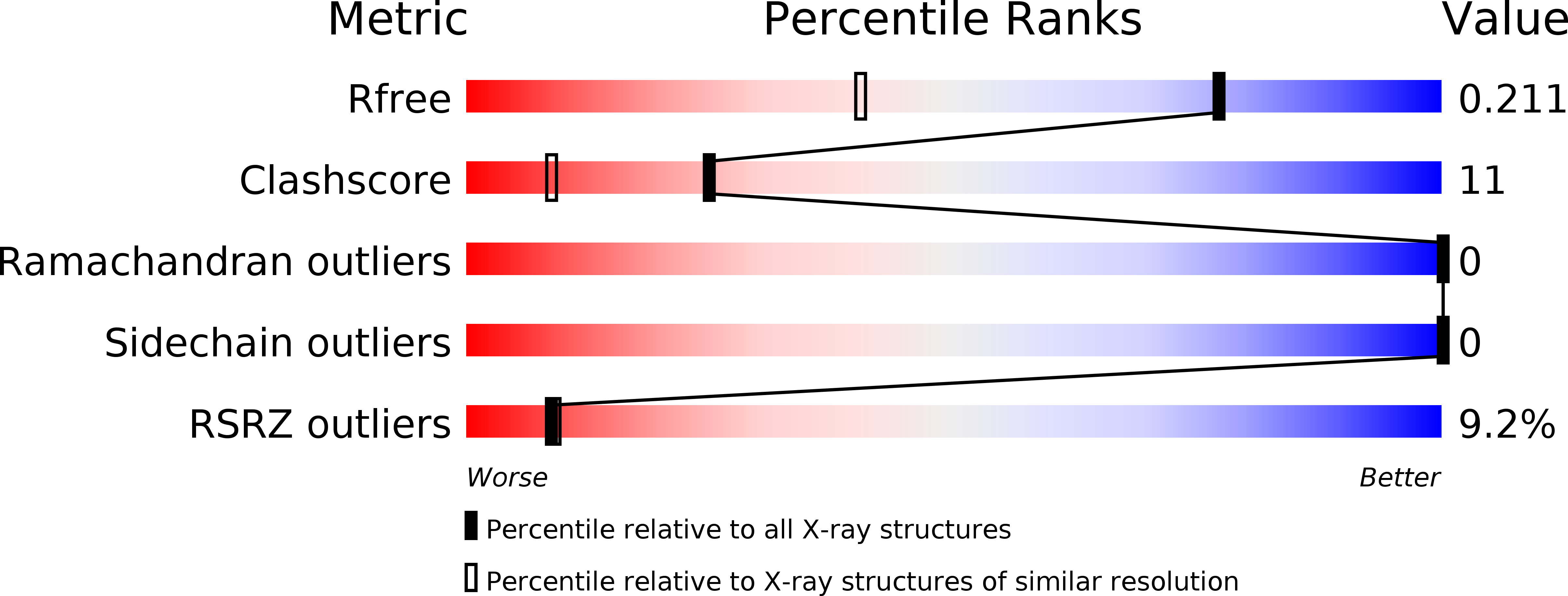
X-ray crystal structure of Corynebacterium glutamicum Nrdh-redoxin at 1.5A
Biological Source:
Source Organism:
Host Organism:
Method Details:
Experimental Method:
Resolution:
1.50 Å
R-Value Free:
0.21
R-Value Work:
0.19
R-Value Observed:
0.19
Space Group:
P 65 2 2
Macromolecular Entities
Structures with similar UniProt ID
Protein Blast
Polymer Type:polypeptide(L)
Molecule:Putative glutaredoxin NrdH
Gene (Uniprot):nrdH
Chain IDs:A
Chain Length:77
Number of Molecules:1
Biological Source:Corynebacterium glutamicum
Primary Citation
NrdH-redoxin of Mycobacterium tuberculosis and Corynebacterium glutamicum Dimerizes at High Protein Concentration and Exclusively Receives Electrons from Thioredoxin Reductase.
J.Biol.Chem. 288 7942 7955 (2013)
PMID: 23362277 DOI: 10.1074/jbc.M112.392688

Abstact

NrdH-redoxins are small reductases with a high amino acid sequence similarity with glutaredoxins and mycoredoxins but with a thioredoxin-like activity. They function as the electron donor for class Ib ribonucleotide reductases, which convert ribonucleotides into deoxyribonucleotides. We solved the x-ray structure of oxidized NrdH-redoxin from Corynebacterium glutamicum (Cg) at 1.5 Å resolution. Based on this monomeric structure, we built a homology model of NrdH-redoxin from Mycobacterium tuberculosis (Mt). Both NrdH-redoxins have a typical thioredoxin fold with the active site CXXC motif located at the N terminus of the first α-helix. With size exclusion chromatography and small angle x-ray scattering, we show that Mt_NrdH-redoxin is a monomer in solution that has the tendency to form a non-swapped dimer at high protein concentration. Further, Cg_NrdH-redoxin and Mt_NrdH-redoxin catalytically reduce a disulfide with a specificity constant 1.9 × 10(6) and 5.6 × 10(6) M(-1) min(-1), respectively. They use a thiol-disulfide exchange mechanism with an N-terminal cysteine pKa lower than 6.5 for nucleophilic attack, whereas the pKa of the C-terminal cysteine is ~10. They exclusively receive electrons from thioredoxin reductase (TrxR) and not from mycothiol, the low molecular weight thiol of actinomycetes. This specificity is shown in the structural model of the complex between NrdH-redoxin and TrxR, where the two surface-exposed phenylalanines of TrxR perfectly fit into the conserved hydrophobic pocket of the NrdH-redoxin. Moreover, nrdh gene deletion and disruption experiments seem to indicate that NrdH-redoxin is essential in C. glutamicum.

Legend

Protein

Chemical

Disease

Primary Citation of related structures
Feedback Form
Name
Email
Institute
Feedback