4FIO image
Deposition Date 2012-06-10
Release Date 2013-07-24
Last Version Date 2023-09-13
Entry Detail
PDB ID:
4FIO
Keywords:
Title:
Crystal Structure of Methenyltetrahydromethanopterin Cyclohydrolase from Methanobrevibacter ruminantium
Biological Source:
Source Organism:
Host Organism:
Method Details:
Experimental Method:
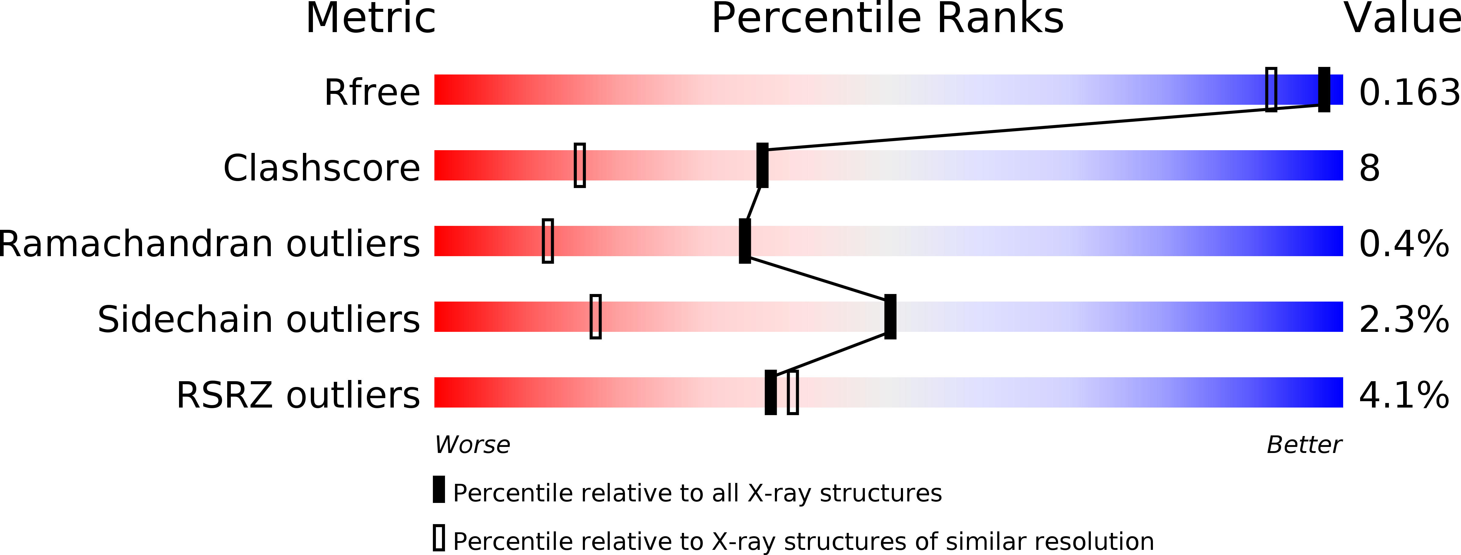
Resolution:
1.37 Å
R-Value Free:
0.17
R-Value Work:
0.13
R-Value Observed:
0.13
Space Group:
P 1
Macromolecular Entities
Polymer Type:polypeptide(L)
Molecule:Methenyltetrahydromethanopterin cyclohydrolase
Gene (Uniprot):mch
Chain IDs:A, B, C
Chain Length:354
Number of Molecules:3
Biological Source:Methanobrevibacter ruminantium
Primary Citation
The crystal structure of methenyltetrahydromethanopterin cyclohydrolase from Methanobrevibacter ruminantium.
Proteins 81 2064 2070 (2013)
PMID: 23873651 DOI: 10.1002/prot.24372

Abstact

Methenyltetrahydromethanopterin cyclohydrolase (Mch) is involved in the methanogenesis pathway of archaea as a C1 unit carrier where N(5) -formyl-tetrahydromethanopterin is converted to methenyl-tetrahydromethanopterin. Mch from Methanobrevibacter ruminantium was cloned, purified, crystallized and its crystal structure solved at 1.37 Å resolution. A biologically active trimer, the enzyme is composed of two domains including an N-terminal domain of six α-helices encompassing a series of four β-sheets and a predominantly anti-parallel β-sheet at the C-terminus flanked on one side by α-helices. Sequence and structural alignments have helped identify residues involved in substrate binding and trimer formation.

Legend

Protein

Chemical

Disease

Primary Citation of related structures