
Deposition Date
2012-06-08
Release Date
2013-06-12
Last Version Date
2023-09-13
Entry Detail
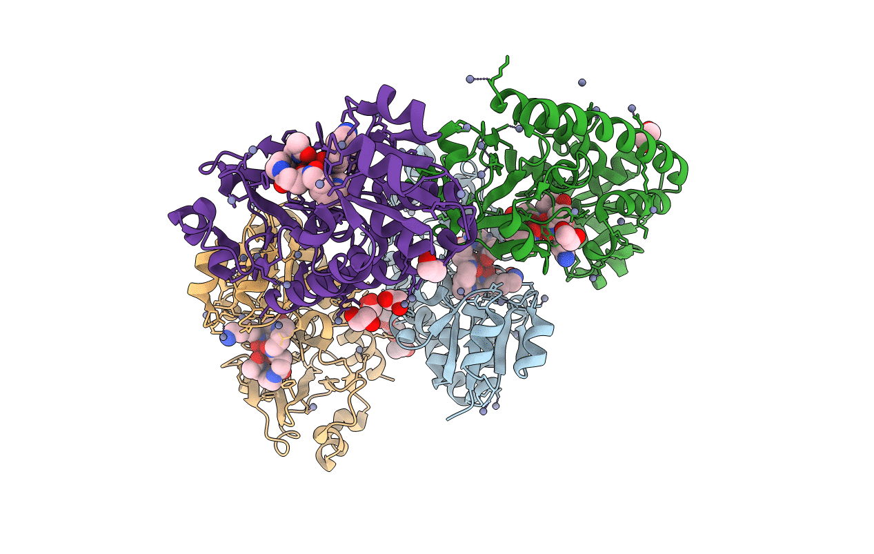
PDB ID:
4FIL
Keywords:
Title:
Structure of FhuD2 from Staphylococcus Aureus with Bound Ferrioxamine B
Biological Source:
Source Organism(s):
Staphylococcus aureus (Taxon ID: 1280)
Expression System(s):
Method Details:
Experimental Method:
Resolution:
2.40 Å
R-Value Free:
0.22
R-Value Work:
0.19
R-Value Observed:
0.20
Space Group:
P 1 21 1
