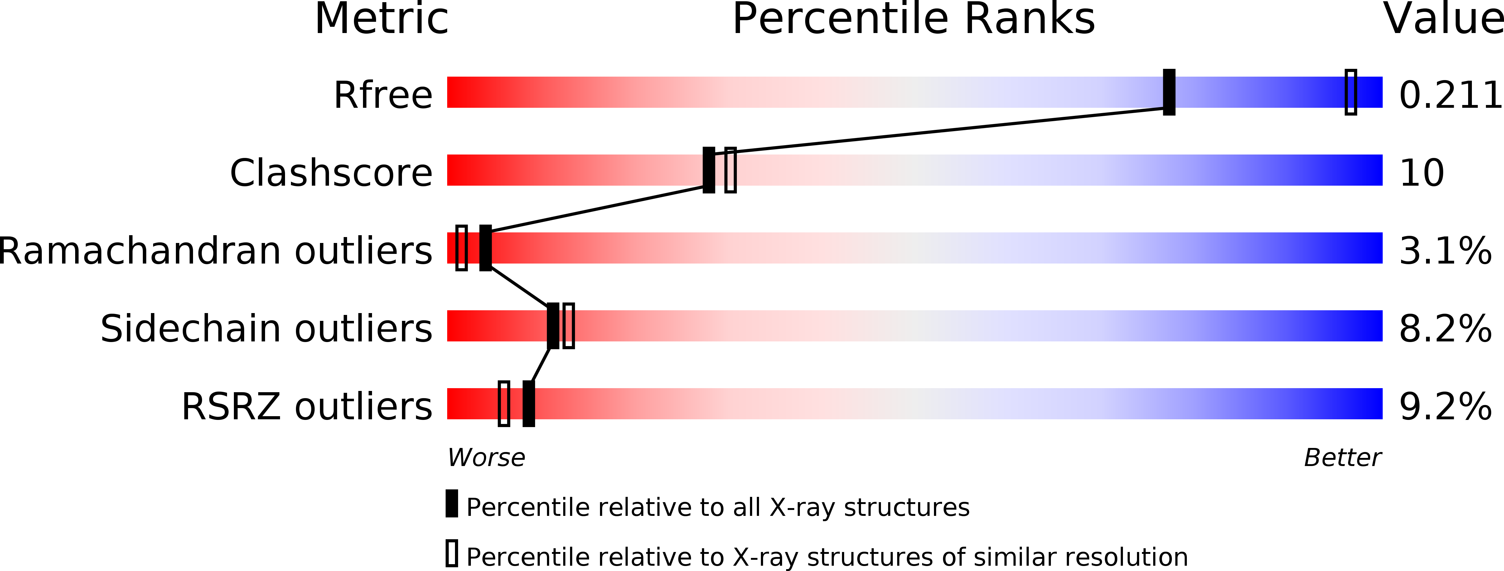
4FGW image
Deposition Date 2012-06-05
Release Date 2012-11-07
Last Version Date 2023-09-13
Entry Detail
PDB ID:
4FGW
Keywords:
Title:
Structure of Glycerol-3-Phosphate Dehydrogenase, GPD1, from Sacharomyces Cerevisiae
Biological Source:
Source Organism:
Host Organism:
Method Details:
Experimental Method:
Resolution:
2.45 Å
R-Value Free:
0.20
R-Value Work:
0.19
R-Value Observed:
0.20
Space Group:
P 43
Macromolecular Entities
Structures with similar UniProt ID
Protein Blast
Polymer Type:polypeptide(L)
Molecule:Glycerol-3-phosphate dehydrogenase [NAD(+)] 1
Gene (Uniprot):GPD1
Chain IDs:A, B
Chain Length:391
Number of Molecules:2
Biological Source:Saccharomyces cerevisiae
Primary Citation
Structure of glycerol-3-phosphate dehydrogenase (GPD1) from Saccharomyces cerevisiae at 2.45A resolution
Acta Crystallogr.,Sect.F 68 1279 1283 (2012)
PMID: 23143232 DOI: 10.1107/S1744309112037736

Abstact

The interconversion of glycerol 3-phosphate and dihydroxyacetone phosphate by glycerol-3-phosphate dehydrogenases provides a link between carbohydrate and lipid metabolism and provides Saccharomyces cerevisiae with protection against osmotic and anoxic stress. The first structure of a glycerol-3-phosphate dehydrogenase from S. cerevisiae, GPD1, is reported at 2.45 Å resolution. The asymmetric unit contains two monomers, each of which is organized with N- and C-terminal domains. The N-terminal domain contains a classic Rossmann fold with the (β-α-β-α-β)2 motif typical of many NAD+-dependent enzymes, while the C-terminal domain is mainly α-helical. Structural and phylogenetic comparisons reveal four main structure types among the five families of glycerol-3-phosphate and glycerol-1-phosphate dehydrogenases and reveal that the Clostridium acetobutylican protein with PDB code 3ce9 is a glycerol-1-phosphate dehydrogenase.

Legend

Protein

Chemical

Disease

Primary Citation of related structures