4FG8 image
Deposition Date 2012-06-04
Release Date 2013-01-23
Last Version Date 2023-09-13
Entry Detail
PDB ID:
4FG8
Keywords:
Title:
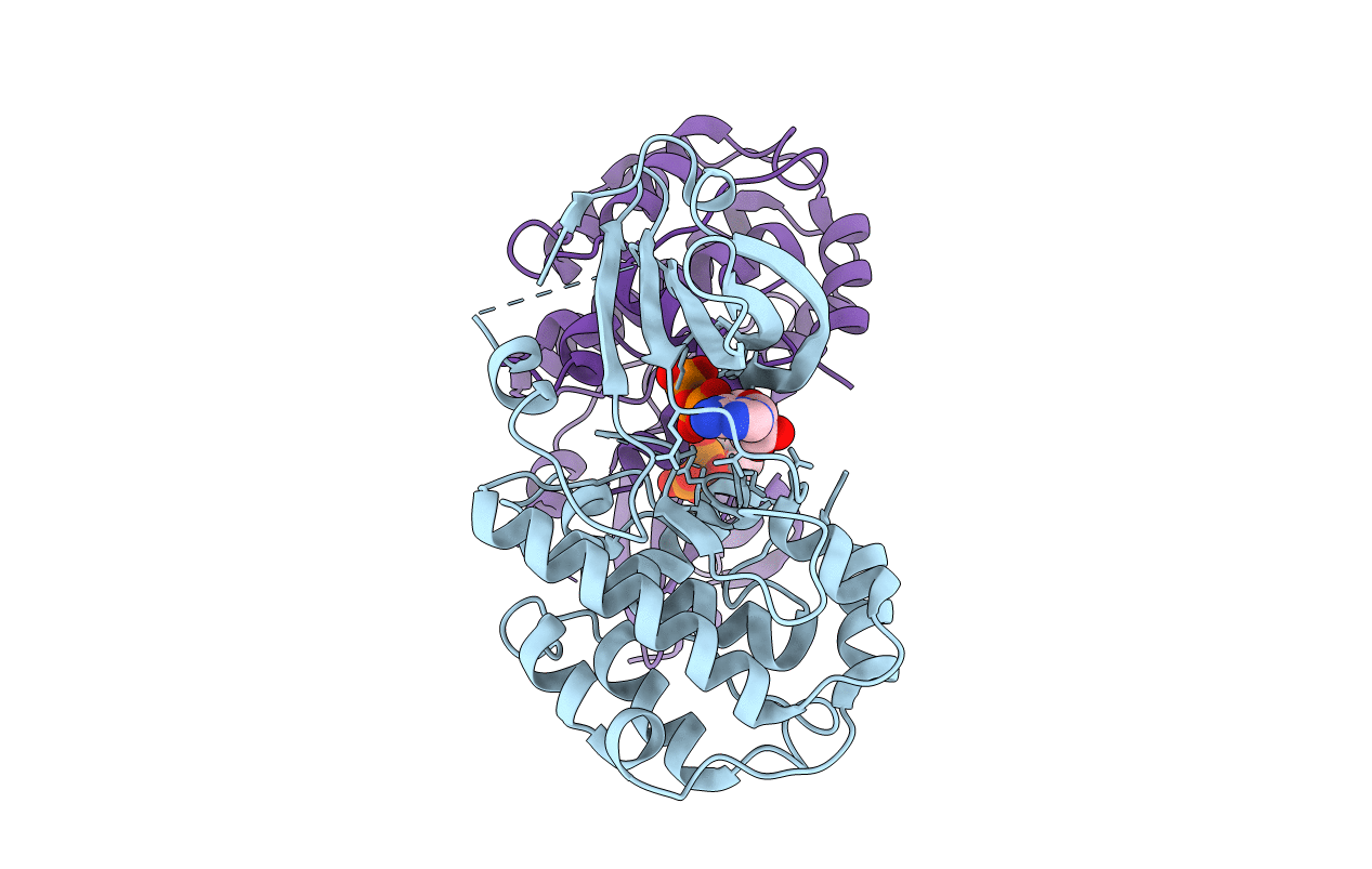
Crystal structure of human calcium/calmodulin-dependent protein kinase I 1-315 in complex with ATP
Biological Source:
Source Organism:
Homo sapiens (Taxon ID: 9606)
Host Organism:
Method Details:
Experimental Method:
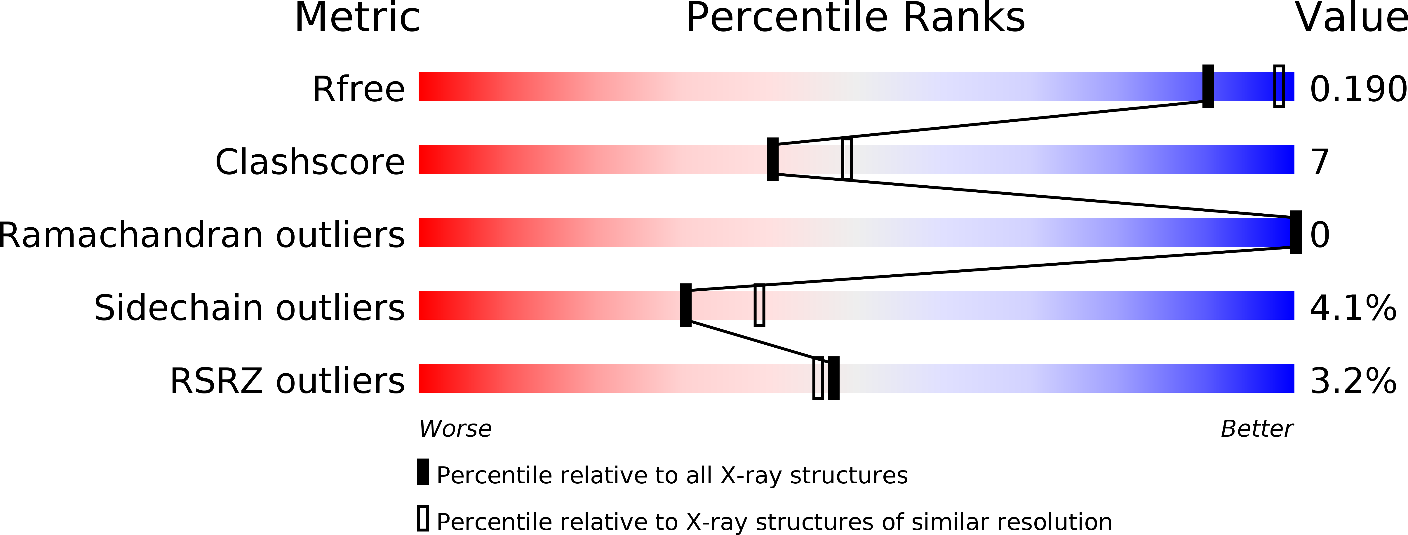
Resolution:
2.20 Å
R-Value Free:
0.24
R-Value Work:
0.19
R-Value Observed:
0.19
Space Group:
P 63
Macromolecular Entities
Polymer Type:polypeptide(L)
Molecule:Calcium/calmodulin-dependent protein kinase type 1
Gene (Uniprot):CAMK1
Chain IDs:A, B
Chain Length:315
Number of Molecules:2
Biological Source:Homo sapiens
Ligand Molecules
Primary Citation
Crystal structures of human CaMKIalpha reveal insights into the regulation mechanism of CaMKI.
Plos One 7 e44828 e44828 (2012)
PMID: 23028635 DOI: 10.1371/journal.pone.0044828

Abstact

Human calcium/calmodulin-dependent protein kinase I (CaMKI) plays pivotal roles in the nervous system. The activity of human CaMKI is regulated by a regulatory region including an autoinhibitory segment and a CaM-binding segment. We report here four structures of three CaMKIα truncates in apo form and in complexes with ATP. In an apo, autoinhibited structure, the activation segment adopts a unique helical conformation which together with the autoinhibitory segment constrains helices αC and αD in inactive conformations, sequesters Thr177 from being phosphorylated, and occludes the substrate-binding site. In an ATP-bound, inactive structure, the activation segment is largely disordered and the CaM-binding segment protrudes out ready for CaM binding. In an ATP-bound, active structure, the regulatory region is dissociated from the catalytic core and the catalytic site assumes an active conformation. Detailed structural analyses reveal the interplay of the regulatory region, the activation segment, and the nucleotide-binding site in the regulation of CaMKI.

Legend

Protein

Chemical

Disease

Primary Citation of related structures