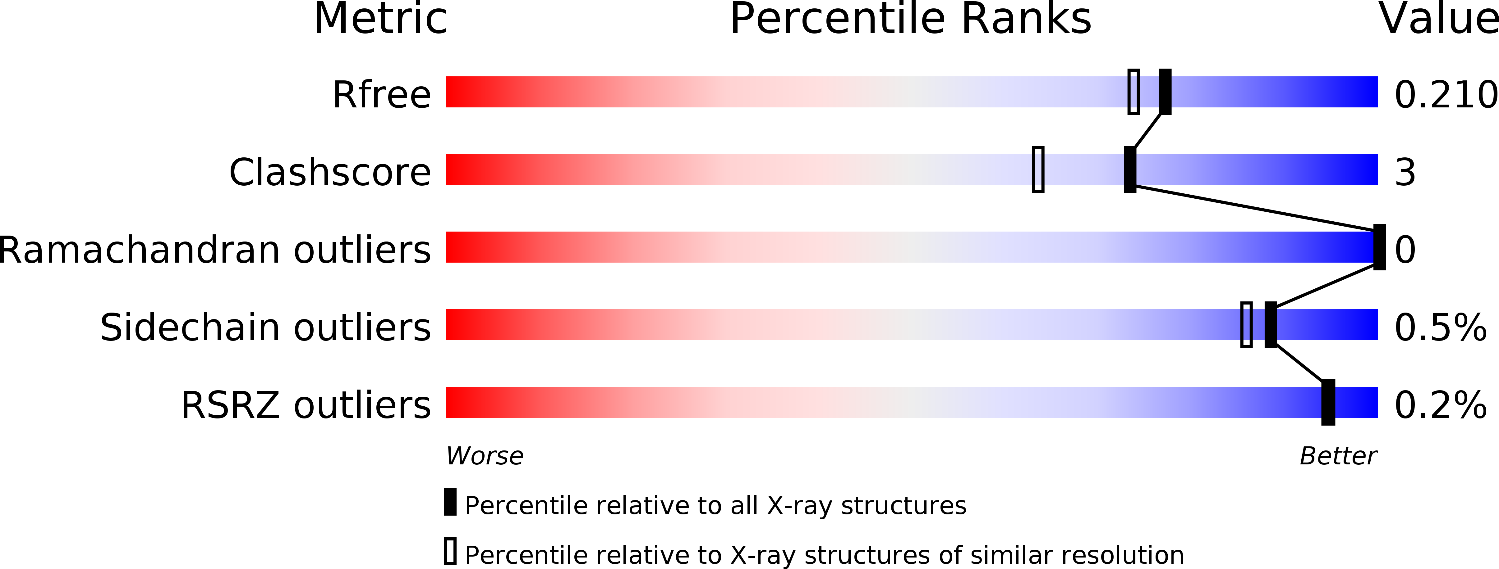
4FF7 image
Deposition Date 2012-05-31
Release Date 2012-08-22
Last Version Date 2023-09-13
Entry Detail
PDB ID:
4FF7
Keywords:
Title:
Structure of C126S mutant of Saccharomyces cerevisiae triosephosphate isomerase
Biological Source:
Source Organism:
Host Organism:
Method Details:
Experimental Method:
Resolution:
1.86 Å
R-Value Free:
0.21
R-Value Work:
0.17
R-Value Observed:
0.17
Space Group:
P 21 21 21
Macromolecular Entities
Structures with similar UniProt ID
Protein Blast
Polymer Type:polypeptide(L)
Molecule:Triosephosphate isomerase
Gene (Uniprot):TPI1
Mutagens:C126S
Chain IDs:A, B
Chain Length:248
Number of Molecules:2
Biological Source:Saccharomyces cerevisiae
Primary Citation
Effects of a buried cysteine-to-serine mutation on yeast triosephosphate isomerase structure and stability.
Int J Mol Sci 13 10010 10021 (2012)
PMID: 22949845 DOI: 10.3390/ijms130810010

Abstact

All the members of the triosephosphate isomerase (TIM) family possess a cystein residue (Cys126) located near the catalytically essential Glu165. The evolutionarily conserved Cys126, however, does not seem to play a significant role in the catalytic activity. On the other hand, substitution of this residue by other amino acid residues destabilizes the dimeric enzyme, especially when Cys is replaced by Ser. In trying to assess the origin of this destabilization we have determined the crystal structure of Saccharomyces cerevisiae TIM (ScTIM) at 1.86 Å resolution in the presence of PGA, which is only bound to one subunit. Comparisons of the wild type and mutant structures reveal that a change in the orientation of the Ser hydroxyl group, with respect to the Cys sulfhydryl group, leads to penetration of water molecules and apparent destabilization of residues 132-138. The latter results were confirmed by means of Molecular Dynamics, which showed that this region, in the mutated enzyme, collapses at about 70 ns.

Legend

Protein

Chemical

Disease

Primary Citation of related structures